Review
. 2022 Feb 10;11(4):613.
doi: 10.3390/cells11040613.

Chromatoid Bodies in the Regulation of Spermatogenesis: Novel Role of GRTH

Affiliations
Review

Chromatoid Bodies in the Regulation of Spermatogenesis: Novel Role of GRTH

Rajakumar Anbazhagan et al. Cells. .

Abstract

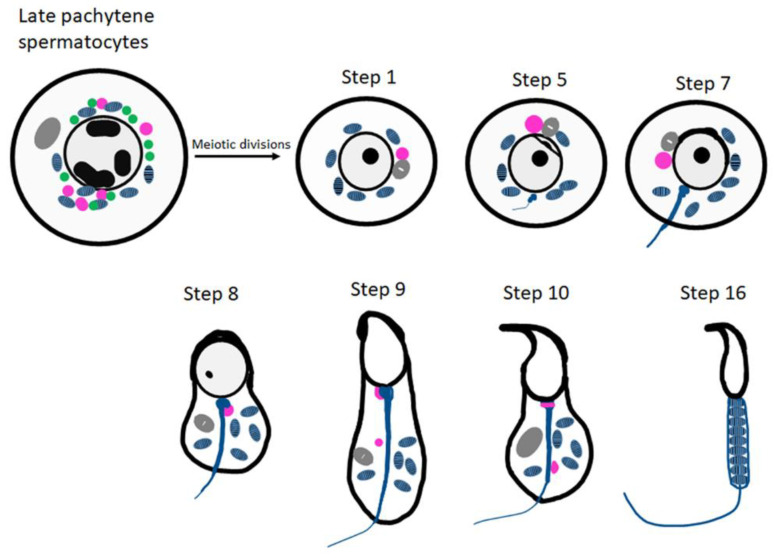
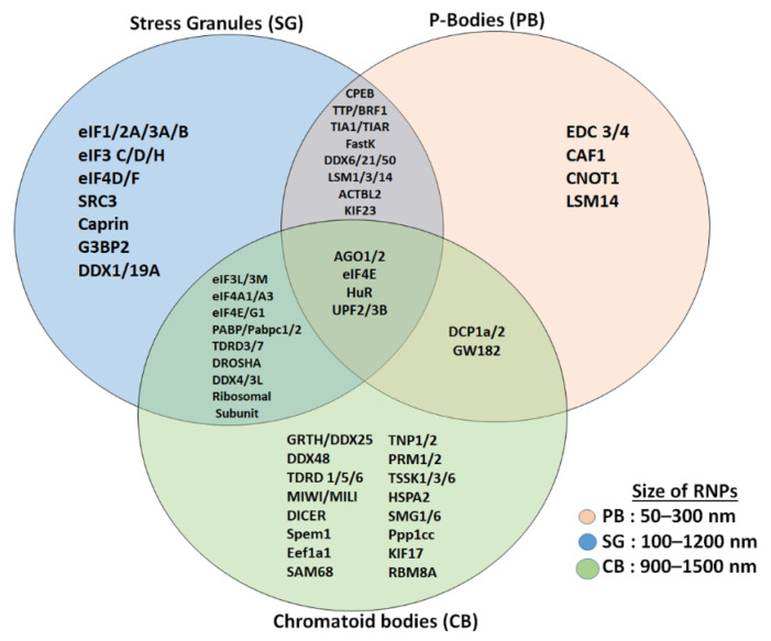
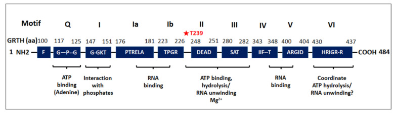
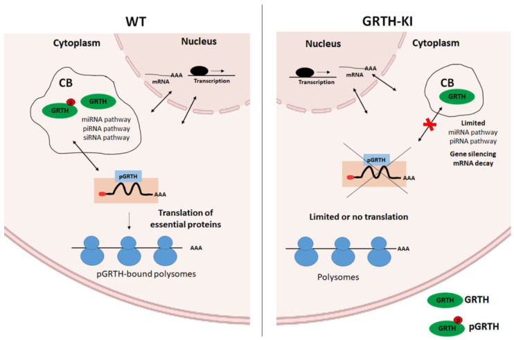
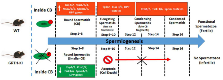
Post-transcriptional and translational control of specialized genes play a critical role in the progression of spermatogenesis. During the early stages, mRNAs are actively transcribed and stored, temporarily bound to RNA binding proteins in chromatoid bodies (CBs). CBs are membrane-less dynamic organelles which serve as storehouses and processing centers of mRNAs awaiting translation during later stages of spermatogenesis. These CBs can also regulate the stability of mRNAs to secure the correct timing of protein expression at different stages of sperm formation. Gonadotropin-regulated testicular RNA helicase (GRTH/DDX25) is an essential regulator of spermatogenesis. GRTH transports mRNAs from the nucleus to the cytoplasm and phospho-GRTH transports mRNAs from the cytoplasm to the CBs. During spermiogenesis, there is precise control of mRNAs transported by GRTH from and to the CBs, directing the timing of translation of critical proteins which are involved in spermatid elongation and acrosomal development, resulting in functional sperm formation. This chapter presents our current knowledge on the role of GRTH, phospho-GRTH and CBs in the control of spermiogenesis. In addition, it covers the components of CBs compared to those of stress granules and P-bodies.

Keywords: RNA storage; RNA transport; chromatoid bodies; phospho-GRTH; spermatogenesis; transcriptome analysis.


Conflict of interest statement

The authors declare no conflict of interest.

Figures

Figure 1
Figure 1
Schematic representation of CB organization and fate during spermiogenesis in mice. The inter-mitochondrial cement (IMC; green) intermixed with mitochondria (blue) and the CB precursors (pink) co-exist in late pachytene spermatocytes. The Golgi complex is depicted in gray. The CB (pink) is condensed to its final single form in the early round spermatids. At step 8 of spermiogenesis, the CB is found at the basis of the flagellum. Later, it splits into two separate structures (step 9 onwards) and eventually disappears.
Figure 2
Figure 2
Comparison of proteins (mRNA decay and translation machinery) present in CBs with stress granules and P-bodies. Stress granules and CBs share several initiation factors and translation initiation assembly proteins, while P-bodies and CBs share few of the proteins. CBs are more related to stress granules than to P-bodies and CBs are bigger in size compared to stress granules and P-bodies.
Figure 3
Figure 3
Schematic representation of the conserved motifs of the DEAD-box family of RNA helicase present in GRTH. Nine conserved motifs of GRTH protein were indicated as Q, I, Ia, Ib, II-VI. Amino acid positions of GRTH corresponding to each conserved motif are indicated on top of each motif. Aside from the conserved motifs, GRTH displays low amino acid similarity with other DEAD-box family of RNA helicases. Amino acids within the conserved motifs are specified by their letter code. Among the several serine and threonine residues in the GRTH, only T239 is phosphorylated by PKA (which is highlighted by a red asterisk).
Figure 4
Figure 4
Cellular and transcriptomic changes occurring in CBs of GRTH-KI mice compared to wild-type mice. (A) Immunofluorescence staining of MVH/DDX4 (green) in the testicular tissue sections of WT and KI mice, CBs indicated by arrow; scale bar: ~25 μm [10]. (B) EM images from testicular sections with round spermatids (RS) showing a lobular structure with an irregular network of the less dense strands characteristic of the CB clearly visible in WT, while in GRTH-KI, CB is markedly reduced in size. Arrows indicate CBs and N-Nucleus; scale bar: ~1 μm [10]. (C) Immunofluorescence staining of MVH/DDX4 as a marker showing CBs (red) isolated from GRTH-KI mice are smaller in size compared to WT. Inset shows detail of CBs indicated by arrow; scale bar: ~25 μm [10]. (D) Comparison of gene expression of downregulated/upregulated genes in germ cells with a decrease/increase in the abundance of genes in CB obtained from wild-type and GRTH-KI mice [10]. Detailed methodology on immunofluorescence, EM, and RNA-Seq analysis were described earlier [10].
Figure 5
Figure 5
RNA transport and associated changes occurring in CBs of GRTH-KI mice compared to wild-type mice. GRTH transports essential mRNAs from nucleus to the cytoplasm for translation. The pGRTH, through its interaction with actively elongating polyribosomes, regulates translation of target mRNA. It is involved in transport of mRNPs in and out of the chromatoid body, where it is transiently stored and/or degraded when not needed in the CBs. mRNAs are transported from the CB by pGRTH for translation at specific times during spermatogenesis.
Figure 6
Figure 6
Schematic diagram showing progression of the spermatogenesis process in the presence and absence of pGRTH, with reference to chromatoid bodies and the associated mRNA and protein expression. Loss of phospho-GRTH altered the CB structure and biochemical composition. It also diminished the transport of essential transcripts between cytoplasm and CBs, thereby altering the mRNA storage profiles and causing impaired spermatid elongation, resulting in loss of spermatozoa and subsequently infertility.

References

    1. Steger K. Haploid spermatids exhibit translationally repressed mRNAs. Anat. Embryol. 2001;203:323–334. doi: 10.1007/s004290100176. - DOI - PubMed
    1. Kimmins S., Sassone-Corsi P. Chromatin remodelling and epigenetic features of germ cells. Nature. 2005;434:583–589. doi: 10.1038/nature03368. - DOI - PubMed
    1. Griswold M.D. Spermatogenesis: The Commitment to Meiosis. Physiol. Rev. 2016;96:1–17. doi: 10.1152/physrev.00013.2015. - DOI - PMC - PubMed
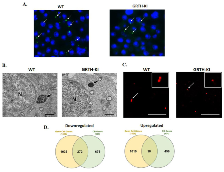
    1. Kavarthapu R., Anbazhagan R., Raju M., Morris C.T., Pickel J., Dufau M.L. Targeted knock-in mice with a human mutation in GRTH/DDX25 reveals the essential role of phosphorylated GRTH in spermatid development during spermatogenesis. Hum. Mol. Genet. 2019;28:2561–2572. doi: 10.1093/hmg/ddz079. - DOI - PMC - PubMed
    1. Shima J.E., McLean D.J., McCarrey J.R., Griswold M.D. The murine testicular transcriptome: Characterizing gene expression in the testis during the progression of spermatogenesis. Biol. Reprod. 2004;71:319–330. doi: 10.1095/biolreprod.103.026880. - DOI - PubMed

Publication types

LinkOut - more resources