Review
. 2022 Feb 15;11(4):672.
doi: 10.3390/cells11040672.

Epigenetic Regulation of Cellular Senescence

Affiliations
Review

Epigenetic Regulation of Cellular Senescence

Jack Crouch et al. Cells. .

Abstract

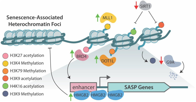
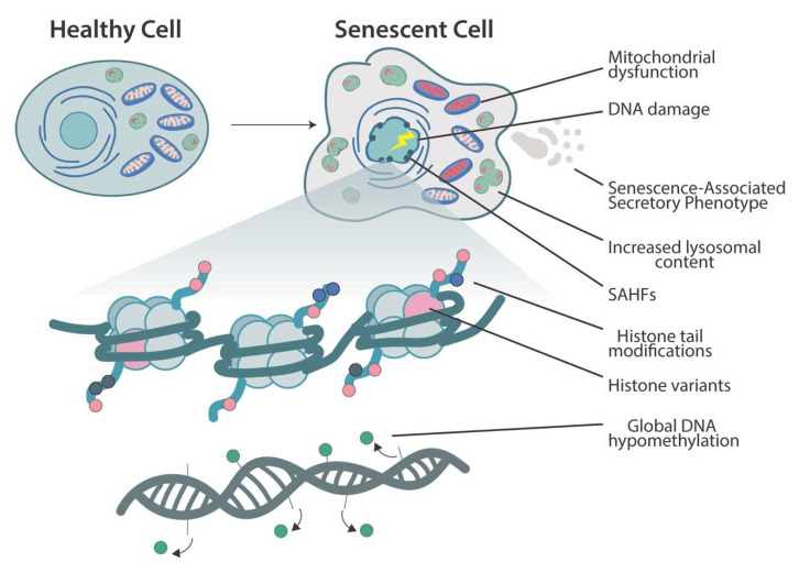
Senescence is a complex cellular stress response that abolishes proliferative capacity and generates a unique secretory pattern that is implicated in organismal aging and age-related disease. How a cell transitions to a senescent state is multifactorial and often requires transcriptional regulation of multiple genes. Epigenetic alterations to DNA and chromatin are powerful regulators of genome architecture and gene expression, and they play a crucial role in mediating the induction and maintenance of senescence. This review will highlight the changes in chromatin, DNA methylation, and histone alterations that establish and maintain cellular senescence, alongside the specific epigenetic regulation of the senescence-associated secretory phenotype (SASP).

Keywords: DNA methylation; SASP; aging; epigenetics; histone modification; senescence.


Conflict of interest statement

The authors declare no conflict of interest.

Figures

Figure 2
Figure 2
An overview of the histone acetylation/methylation marks and proteins that regulate genes involved in the senescence-associated secretory phenotype (SASP). High-mobility-group protein B2 (HMGB2) binds loci of key SASP genes preventing their incorporation into the SAHF during senescence allowing for their transcription [120]. Mixed-lineage leukemia 1 (MLL1) methylates H3 at Lys-4 to promote transcription of SASP genes [122]. Disruptor of telomeric silencing 1-like (DOT1L) mediates methylation of H3 at Lys-79 driving transcription of potent inflammatory cytokine and SASP factor IL1A [123]. Bromodomain-containing protein 4 (BRD4) mediated acetylation of H3 at Lys-27 is necessary to drive transcription of SASP factors during senescence [121]. G9a and G9a-like proteins, which deposit repressive methylation on H3 at Lys-9, prevent transcription of SASP genes in proliferating cells [124]. In response to DNA damage, these proteins are degraded, allowing for active transcription of IL-6 and IL-8. NAD-dependent deacetylase sirtuin-1 (SIRT-1) negatively regulates SASP expression through deacetylation of H4 Lys-16 and H3 Lys-9 in normal proliferating cells. Senescence induction decreases expression of SIRT-1, allowing for acetylation and expression of key SASP genes [126].
Figure 1
Figure 1
An overview of the morphological, cellular, and epigenomic changes that occur as cell senescence. Morphological alterations include cellular flattening and enlargement. Cellular lysosomal content increases concomitantly with the elevated expression of senescence-associated β-galactosidase, a highly specific marker for senescence [19]. Mitochondrial abnormalities include impaired biogenesis and mitophagy along with increased reactive oxygen species production and decreased membrane potential [20]. Senescent cells also secrete a heterogeneous group of proinflammatory cytokines, chemokines, proteases, and growth factors that have a profound impact on neighboring cells, known as senescence-associated secretory phenotype (SASP). Large-scale chromatin reorganization occurs with the generation of senescence-associated heterochromatin foci (SAHF), which specifically suppress transcription of pro-proliferation genes [23]. Global hypomethylation of DNA is observed in replicative senescence [29]. Histone variant deposition, histone tail acetylation/methylation, and DNA methylation encompass many of the epigenomic alterations that initiate and maintain cellular senescence.

References

    1. López-Otín C., Blasco M.A., Partridge L., Serrano M., Kroemer G. The hallmarks of aging. Cell. 2013;153:1194–1217. doi: 10.1016/j.cell.2013.05.039. - DOI - PMC - PubMed
    1. Campisi J. Cellular senescence as a tumor-suppressor mechanism. Trends Cell Biol. 2001;11:S27–S31. doi: 10.1016/S0962-8924(01)02151-1. - DOI - PubMed
    1. Demaria M., Ohtani N., Youssef S.A., Rodier F., Toussaint W., Mitchell J.R., Laberge R.-M., Vijg J., Van Steeg H., Dollé M.E., et al. An Essential Role for Senescent Cells in Optimal Wound Healing through Secretion of PDGF-AA. Dev. Cell. 2014;31:722–733. doi: 10.1016/j.devcel.2014.11.012. - DOI - PMC - PubMed
    1. Ritschka B., Storer M., Mas A., Heinzmann F., Ortells M.C., Morton J.P., Sansom O.J., Zender L., Keyes W.M. The senescence-associated secretory phenotype induces cellular plasticity and tissue regeneration. Genes Dev. 2017;31:172–183. doi: 10.1101/gad.290635.116. - DOI - PMC - PubMed
    1. Baker D.J., Childs B.G., Durik M., Wijers M.E., Sieben C.J., Zhong J., Saltness R.A., Jeganathan K.B., Verzosa G.C., Pezeshki A., et al. Naturally occurring p16Ink4a-positive cells shorten healthy lifespan. Nature. 2016;530:184–189. doi: 10.1038/nature16932. - DOI - PMC - PubMed

Publication types

LinkOut - more resources