. 2022 Jan 18;10(2):205.
doi: 10.3390/biomedicines10020205.

L-Arginine Depletion Improves Spinal Cord Injury via Immunomodulation and Nitric Oxide Reduction

Affiliations

L-Arginine Depletion Improves Spinal Cord Injury via Immunomodulation and Nitric Oxide Reduction

Céline Erens et al. Biomedicines. .

Abstract

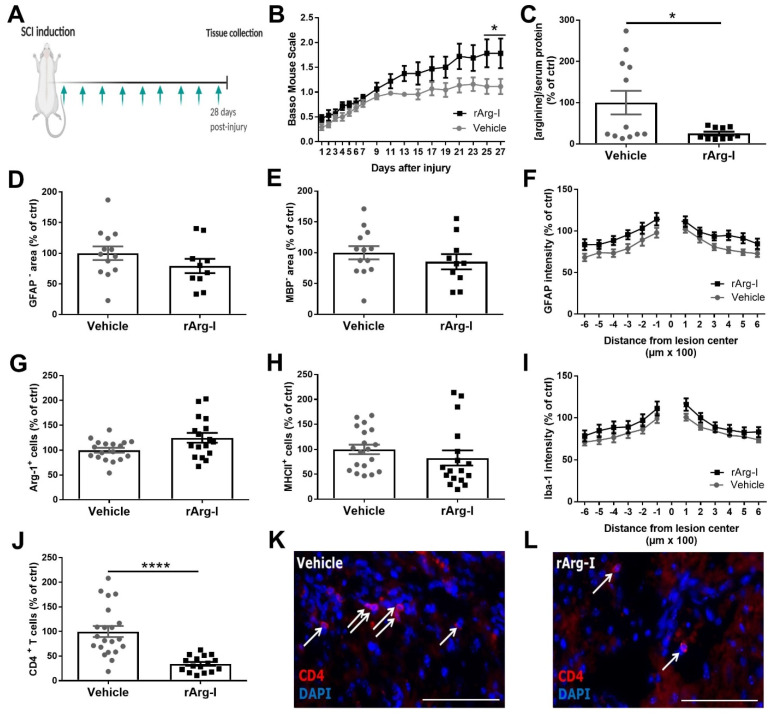
Background: Spinal cord injury (SCI) elicits robust neuroinflammation that eventually exacerbates the initial damage to the spinal cord. L-arginine is critical for the responsiveness of T cells, which are important contributors to neuroinflammation after SCI. Furthermore, L-arginine is the substrate for nitric oxide (NO) production, which is a known inducer of secondary damage.

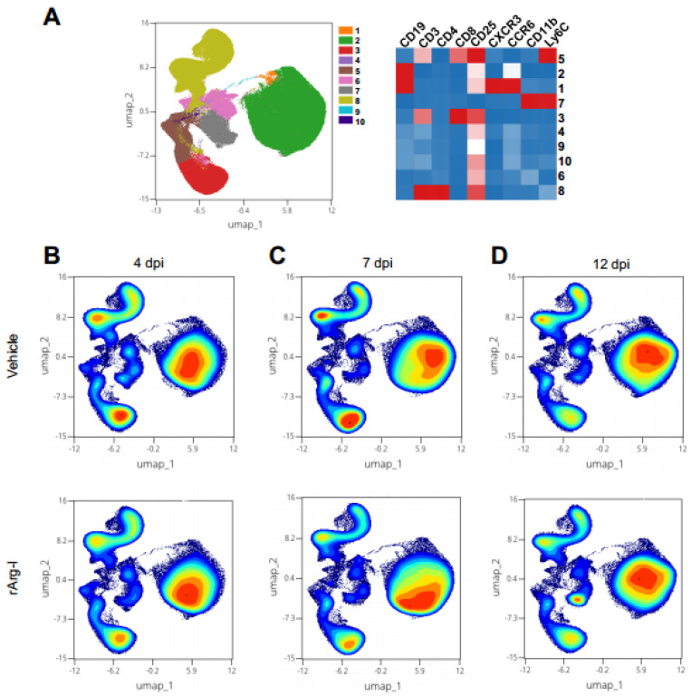
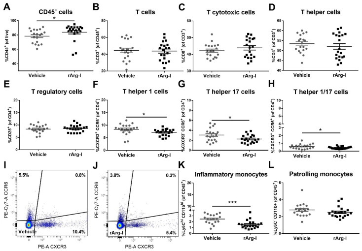
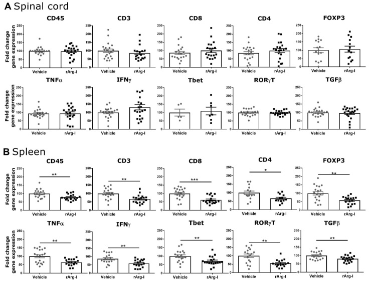
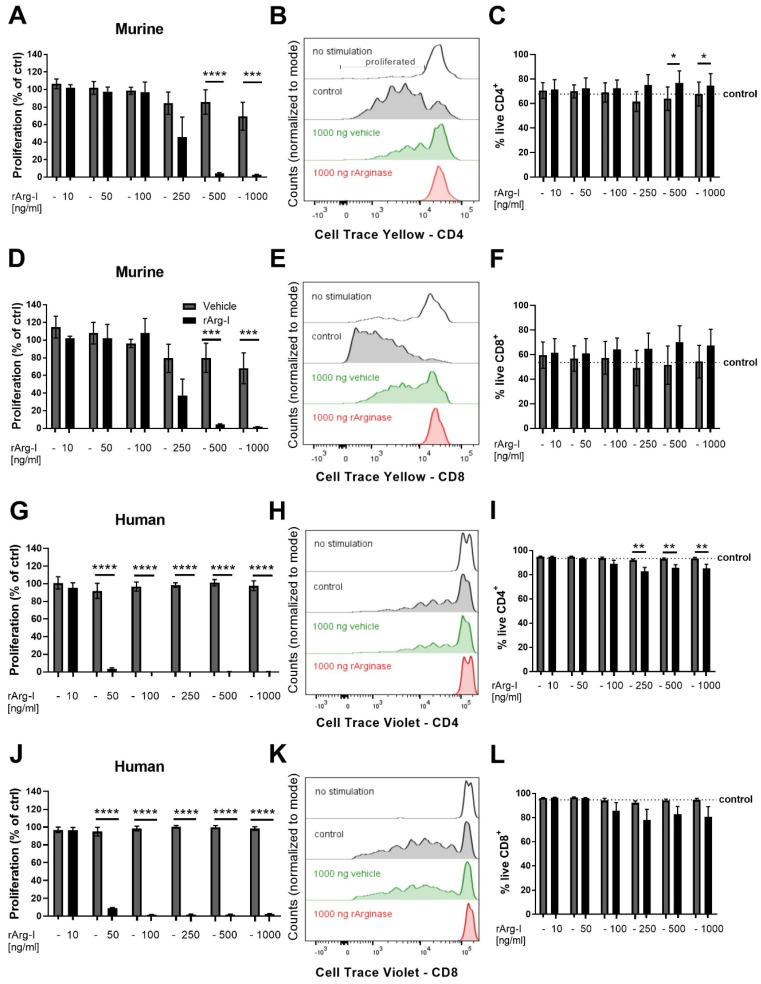
Methods: To accomplish systemic L-arginine depletion, repetitive injections of recombinant arginase-1 (rArg-I) were performed. Functional recovery and histopathological parameters were analyzed. Splenic immune responses were evaluated by flow cytometry. Pro-inflammatory gene expression and nitrite concentrations were measured.

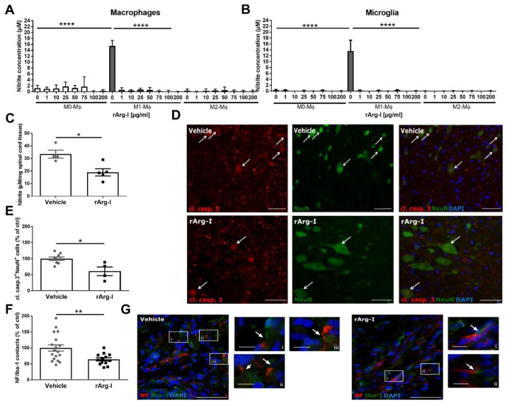
Results: We show for the first time that systemic L-arginine depletion improves locomotor recovery. Flow cytometry and immunohistological analysis showed that intraspinal T-cell infiltration was reduced by 65%, and peripheral numbers of Th1 and Th17 cells were suppressed. Moreover, rArg-I treatment reduced the intraspinal NO production by 40%. Histopathological analyses revealed a 37% and 36% decrease in the number of apoptotic neurons and neuron-macrophage/microglia contacts in the spinal cord, respectively.

Conclusions: Targeting detrimental T-cell responses and NO-production via rArg-I led to a reduced neuronal cell death and an improved functional recovery. These findings indicate that L-arginine depletion holds promise as a therapeutic strategy after SCI.

Keywords: CNS trauma; T cells; arginase-1; neuroinflammation; nitric oxide.


Conflict of interest statement

Hendrix is the founder of smartsciencecareer.com, which does not affect the data presented here. All co-authors of Bio-Cancer treatment International have no conflicts of interest in the present study for publication. P.C. is the founder of Biocancer Treatment International Ltd. P.C., G.S. and S.B. are listed as inventors on a patent (US9789169B2) covering recArg1/BCT-100. The remaining authors declare no competing interests.

Figures

Figure 1
Figure 1
rArg-I treatment improves recovery and reduces intraspinal CD4+ T cell numbers 28 days after trauma. (A) Schematic representation of the treatment protocol. A T-cut hemisection was performed, and rArg-I (50 mg/kg) was injected intraperitoneally every 3 days starting at the day of injury, indicated by the green arrows. Mice were sacrificed 28 days post-injury (dpi). (B) Treatment with rArg-I improved functional recovery after spinal cord injury (SCI). Recovery of hind limb motor function was determined using the Basso Mouse Scale. n = 16–22 mice/group. [F(1,36) = 4.932, p = 0.0115, two-way ANOVA]. (C) Serum arginine depletion was found after rArg-I treatment 28 dpi. n = 10–12 mice/group. [100 ± 28.56 vs. 25.29 ± 4.55, p = 0.0287, two-tailed unpaired student t-test]. (DJ) Histological analyses of spinal cord sections 28 dpi. (D) Lesion size, (E) demyelinated area, (F) astrogliosis, (G,H) number of arg-1+ and MHCII+ cells, and (I) Iba-1 intensity did not change between treatment groups. n = 10–19 mice/group. (J) The number of infiltrating CD4+ T cells was significantly reduced in the spinal cord of mice treated with rArg-I compared to vehicle control 28 dpi. n = 16–21 mice/group. [100 ± 11.09 vs. 34.33 ± 3.98, p < 0.0001, two-tailed unpaired student t-test]. Representative images of CD4+ T cells (white arrows) in (K) vehicle- and (L) rArg-I-treated mice, respectively. Scale bars represent 75 µm. Data are pooled from two independently performed experiments and represent mean ± SEM. * p < 0.05 and **** p < 0.0001.
Figure 2
Figure 2
The splenic immune subsets remain unaltered within the first two weeks after spinal cord injury. (A) The UMAP plot, overlayed with FlowSOM metaclusters, revealed major immune subsets in the spleen, including CD4+ helper and CD8+ cytotoxic T cells, B cells, patrolling and inflammatory monocytes (left). The marker expression (red = high expression, blue = low expression) of these cells is indicated on the heat map (right). The identified cell populations are numbered from 1–10 in the UMAP and heat map, respectively. There were no major shifts in these subsets (B) 4, (C) 7, and (D) 12 dpi identified. Representative plots are shown. n = 18–20 mice/group.
Figure 3
Figure 3
The acute splenic immune response is affected by rArg-I-mediated L-arginine depletion after spinal cord injury. (A) Flow cytometry indicated increased numbers of CD45+ leukocytes 4 dpi when mice were treated with rArg-I. n = 19–20 mice/group. [81.3 vs. 88, p = 0.0115, two-tailed Mann–Whitney U test]. L-arginine depletion had no effect on the number of splenic (B) CD3+ T cells, (C) CD8+ T cytotoxic cells, (D) CD4+ T helper cells, and (E) CD4+CD25+ double positive T regulatory cells 4 dpi. n = 19–20 mice/group. (FH) The number of Th1, Th17, and Th1/17 cells was significantly decreased when rArg-I was applied compared to the vehicle. n = 19–20 mice/group. [p = 0.0431, p = 0.0253, and p = 0.0322, two-tailed unpaired student t-test]. (I,J) Representative dot plots of splenocytes isolated from (I) vehicle-treated and (J) rArg-I-treated mice. (K) rArg-I treatment significantly decreased the percentage of splenic inflammatory monocytes. n = 5–20 mice/group. [2.46 vs. 1.32, p = 0.0003, two-tailed Mann–Whitney U test]. (L) The number of patrolling monocytes remained unaltered 4 dpi. Data are shown as mean ± SEM. * p < 0.05 and *** p < 0.001.
Figure 4
Figure 4
rArg-I treatment decreases the transcription of selected inflammatory genes in the spleen (systemic inflammation). (A) Quantitative RT-PCR analysis of spinal cord tissue isolated 7 days after spinal cord injury (SCI) induction. The expression of all the tested inflammatory genes did not change between treatment groups. n = 5–20 mice/group. (B) Quantitative RT-PCR analysis of spleen samples isolated 7 days after SCI. All analyzed inflammatory genes decreased in the rArg-I treatment group compared to vehicle control. n = 13–19 mice/group. [p < 0.05, p < 0.01, p < 0.001, two-tailed unpaired student t-test and two-tailed Mann–Whitney U test]. Data are shown as mean ± SEM, fold change of vehicle control. * p < 0.05, ** p < 0.01 and *** p < 0.001.
Figure 5
Figure 5
CD4+ and CD8+ murine and human T-cell proliferation is significantly reduced by rArg-I. Doubling rates of murine splenocytes and human peripheral blood mononuclear cells (PBMCs) were analyzed utilizing Cell Trace Yellow and Violet labelling, respectively. Cell viability was checked 5–6 days after the onset of the experiment. (A,D,G,J) The proliferation of murine and human T cells are indicated as percentage of control (no vehicle or rArg-I incubation). (B,E,H,K) Corresponding representative histograms. (A,B) Murine T-cell activation resulted in CD4+ T-cell proliferation. rArg-I addition in concentrations of 500 ng/mL or more, reduced proliferation. n = 5 biological repeats. [F(1,21) = 37.65, p < 0.0001, two-way ANOVA]. (D,E) Supplementation of rArg-I suppressed murine CD8+ T-cell proliferation. n = 5 biological repeats. [F(1,21) = 28.06, p < 0.0001, two-way ANOVA]. (G,H) CD4+ human PBMC proliferation was ablated when rArg-I concentrations equal to or higher than 50 ng/mL were added. n = 5–7 biological repeats. [F(1,30) = 1356, p < 0.0001, two-way ANOVA]. (J,K) A similar reduction was found in the human CD8+ PBMC cell population. n = 5–7 biological repeats. [F(1,30) = 6124, p < 0.0001, two-way ANOVA]. In all conditions, the vehicle solution had no impact on the doubling rate. (C,F) rArg-I treatment did not affect murine T-cell survival. n = 5 biological repeats. (I) Human blood-derived CD4+ T cells showed significantly decreased viability when incubated with rArg-I compared to the vehicle when concentrations higher than 100 ng/mL were applied. n = 5–7 biological repeats. [F(1,30) = 34.63, p < 0.01, two-way ANOVA]. (L) Human CD8+ PBMCs showed no sign of decreased viability for rArg-I. n = 5–7 biological repeats. (C,F,I,L) The mean survival of control samples not exposed to vehicle or rArg-I is indicated by the dotted line. The cell viability is indicated as the percentage of FVD eFluor506-negative cells within the CD4+ or CD8+ T gates, respectively. The vehicle solution had no impact on the cell survival for both murine and human cells. Grey-filled bars are vehicle controls, black-filled bars represent rArg-I-treated cells. Data are shown as mean ± SEM. * p < 0.05, ** p < 0.01, *** p < 0.001, and **** p < 0.0001.
Figure 6
Figure 6
L-arginine depletion through rArg-I supplementation reduces NO production, neurotoxicity, and the number of phagocyte/axon contacts. (A) The NO production of bone marrow-derived macrophages exposed to inflammatory stimuli (M1-Mϕ) was significantly attenuated when rArg-I was added to the culture media (added concentrations indicated below graph). As a negative control, non-polarized (M0-Mϕ) and rIL-13 incubated macrophages (M2-Mϕ) were used. n = 5–9 biological repeats. [F(23,112) = 26.81, p < 0.0001, one-way ANOVA]. (B) Griess assay showed similar results on primary microglia; supplementation of rArg-I significantly decreases the nitrite concentration in the presence of inflammatory stimuli. n = 4–5 biological repeats. [F(23,83) = 11.1, p < 0.0001, one-way ANOVA]. (C) The perilesional nitrite concentration in spinal cord homogenates was significantly reduced 7 days post-injury (dpi) in rArg-I-treated animals compared to vehicle controls. n = 4–5 mice/group. [31.54 vs. 18.56, p = 0.0317, two-tailed Mann–Whitney test]. (D,E) Histological analysis and representative images of the cleaved caspase 3/NeuN staining of spinal cord tissue from spinal cord injury (SCI) animals treated with vehicle solution or rArg-I, respectively. A marked reduction in the number of cleaved caspase 3+ NeuN+ double positive cells (white arrows) is observed 28 dpi in rArg-I-treated mice. n = 4–9 mice/group. [98.85 vs. 61.84, p = 0.0336, two-tailed Mann–Whitney test]. (F,G) Quantification and representative pictures of perilesional macrophage/microglia and axon contacts 28 dpi. Treatment with rArg-I significantly reduced the number of contacts between Iba1+ and NF+ cells (white boxed regions). The white boxed regions (iiii) in the micrographs are shown in a higher magnification to indicate examples of microglia/macrophage and axon contacts. Data are pooled from two independently performed experiments. n = 13–18 mice/group. [100 ± 9.923 vs. 64.32 ± 4.923, p = 0.0077, two-tailed unpaired student t-test]. (D,G) Scale bars represent 50 µm, for the magnified pictures i–iii 10 µm. Data represent mean ± SEM. Cl. Casp. 3: cleaved caspase 3. * p < 0.05, ** p < 0.01, **** p < 0.0001.

Similar articles

Cited by

References

    1. Rouanet C., Reges D., Rocha E., Gagliardi V., Silva G.S. Traumatic spinal cord injury: Current concepts and treatment update. Arq. Neuropsiquiatr. 2017;75:387–393. doi: 10.1590/0004-282x20170048. - DOI - PubMed
    1. Beck K.D., Nguyen H.X., Galvan M.D., Salazar D.L., Woodruff T.M., Anderson A.J. Quantitative analysis of cellular inflammation after traumatic spinal cord injury: Evidence for a multiphasic inflammatory response in the acute to chronic environment. Brain. 2010;133 (Pt. 2):433–447. doi: 10.1093/brain/awp322. - DOI - PMC - PubMed
    1. Kigerl K.A., McGaughy V.M., Popovich P.G. Comparative analysis of lesion development and intraspinal inflammation in four strains of mice following spinal contusion injury. J. Comp. Neurol. 2006;494:578–594. doi: 10.1002/cne.20827. - DOI - PMC - PubMed
    1. Popovich P.G., Wei P., Stokes B.T. Cellular inflammatory response after spinal cord injury in Sprague-Dawley and Lewis rats. J. Comp. Neurol. 1997;377:443–464. doi: 10.1002/(SICI)1096-9861(19970120)377:3<443::AID-CNE10>3.0.CO;2-S. - DOI - PubMed
    1. Sroga J.M., Jones T.B., Kigerl K.A., McGaughy V.M., Popovich P.G. Rats and mice exhibit distinct inflammatory reactions after spinal cord injury. J. Comp. Neurol. 2003;462:223–240. doi: 10.1002/cne.10736. - DOI - PubMed