. 2022 Jan 18;10(2):201.
doi: 10.3390/biomedicines10020201.

VEGF-Trap Modulates Retinal Inflammation in the Murine Oxygen-Induced Retinopathy (OIR) Model

Affiliations

VEGF-Trap Modulates Retinal Inflammation in the Murine Oxygen-Induced Retinopathy (OIR) Model

Jesús Eduardo Rojo Arias et al. Biomedicines. .

Abstract

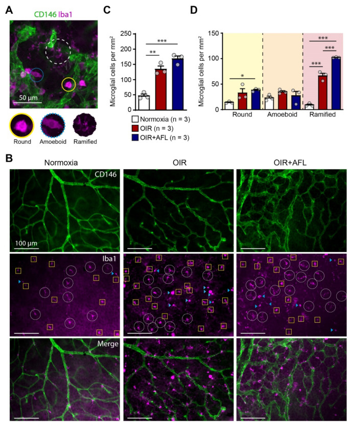
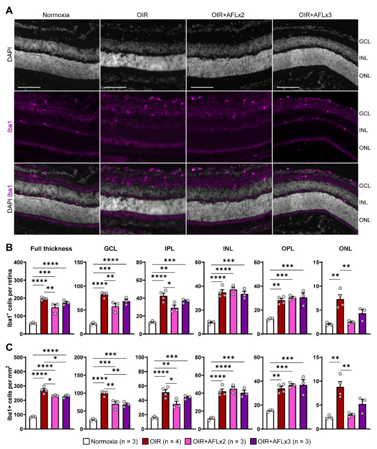
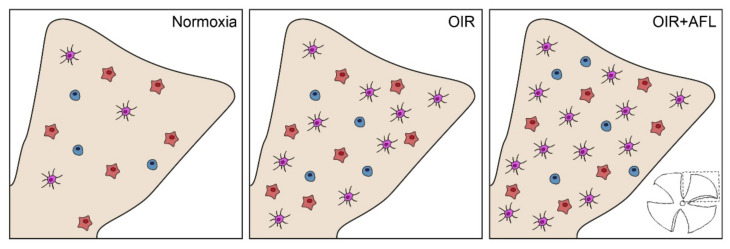
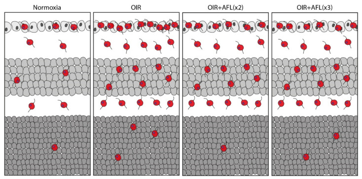
Anti-Vascular Endothelial Growth Factor (VEGF) agents are the first-line treatment for retinal neovascular diseases, which represent the most prevalent causes of acquired vision loss world-wide. VEGF-Trap (Aflibercept, AFL), a recombinant decoy receptor recognizing ligands of both VEGFR-1 and -2, was recently reported to be highly efficient in improving visual acuity and preserving retinal anatomy in individuals affected by diabetic macular edema. However, the precise molecular and cell biological mechanisms underlying the beneficial effects of this novel tool have yet to be elucidated. Using the mouse oxygen-induced retinopathy (OIR) model as a surrogate of retinopathies with sterile post-ischemic inflammation, such as late proliferative diabetic retinopathy (PDR), retinopathy of prematurity (ROP), and diabetic macular edema (DME), we provide evidence that AFL modulates inflammation in response to hypoxia by regulating the morphology of microglial cells, a parameter commonly used as a proxy for changes in their activation state. We show that AFL administration during the hypoxic period of OIR leads to an increased number of ramified Iba1+ microglial cells/macrophages while subsequently limiting the accumulation of these cells in particular retinal layers. Our results suggest that, beyond its well-documented beneficial effects on microvascular regeneration, AFL might exert important modulatory effects on post-ischemic retinal inflammation.

Keywords: Aflibercept; OIR; VEGF-Trap; inflammation; microglia; oxygen-induced retinopathy; retina.


Conflict of interest statement

The authors declare no conflict of interest. The funders had no role in the design of the study; in the collection, analyses, or interpretation of data; in the writing of the manuscript; or in the decision to publish the results.

Figures

Figure 1
Figure 1
The proportion of ramified macrophage/microglial cells increases in retinas from OIR mice upon AFL treatment. (A) Representative micrograph of a CD146 (green) and Iba1 (magenta) immuno-fluorescently labeled retina from a P17 OIR+AFL mouse. CD146-positive structures in the image are neovascular tufts. Iba1+ cells were morphologically discriminated into round (solid line, yellow), amoeboid (dotted line, blue), and ramified (dashed line, white). Scale bar: 50 μm. (BD) Iba1+ cells were quantified according to their morphology in immuno-fluorescently labeled flat-mounted retinas from P17 normoxic (white), OIR (red) and OIR+AFL (blue) mice (n = 3 per treatment). Scale bar: 100 μm. In (B), ramified cells are encircled by a white dashed line, round cells are labelled with a blue arrowhead, and amoeboid cells are enclosed in yellow squares. The images are representative of vascularized regions free of neovascular tufts. In graphs, the total number of Iba1+ cells (C), irrespective of their morphology, as well as the number of cells exhibiting each morphology (D), are shown as quantified in entire retinal flat-mounts. Error bars display +1 S.E.M. n.s., non-significant; * p < 0.05; ** p < 0.01, *** p < 0.001.
Figure 2
Figure 2
Immunofluorescent labeling and quantification of Iba1+ cells in retinal cross sections. (A) Representative micrographs of Iba1 (magenta) immuno-fluorescently labeled retinal cross-sections from P19 Normoxic, OIR, OIR+AFLx2, and OIR+AFLx3 mice. DAPI counterstain is shown in greyscale. GCL, ganglion cell layer; INL, inner nuclear layer; ONL, outer nuclear layer. Scale bar: 100 µm. (B,C) Total numbers (B) and density (C) of Iba1+ cells in the full retina and in distinct retinal layers. IPL, inner plexiform layer; OPL, outer plexiform layer. Error bars display +1 S.E.M. * p < 0.05; ** p < 0.01, *** p < 0.001, **** p < 0.0001.
Figure 3
Figure 3
Schematic representation of Iba1+ cells in P17 flat-mounted normoxic, OIR and OIR+AFL retinas. Round cells are shown in blue, amoeboid in orange, and ramified in purple. One of the four quadrants in which retinal tissues were cut for flat-mounting (bottom right) is schematically shown in beige for each condition (Normoxia, OIR and OIR+AFL).
Figure 4
Figure 4
Schematic representation of Iba1+ cells in P19 cross-sections of normoxic, OIR, OIR+AFLx2, and OIR+AFLx3 retinas. Retinal tissue layers are shown in the following order: ganglion cell layer (GCL), top; inner nuclear layer (INL), middle; outer nuclear layer (ONL), bottom.

Similar articles

Cited by

References

    1. Lee P., Wang C.C., Adamis A.P. Ocular Neovascularization: An Epidemiologic Review. Surv. Ophthalmol. 1998;43:245–269. doi: 10.1016/S0039-6257(98)00035-6. - DOI - PubMed
    1. Chen J., Smith L.E. Retinopathy of prematurity. Angiogenesis. 2007;10:133–140. doi: 10.1007/s10456-007-9066-0. - DOI - PubMed
    1. Hartnett M.E., Penn J.S. Mechanisms and Management of Retinopathy of Prematurity. N. Engl. J. Med. 2012;367:2515–2526. doi: 10.1056/NEJMra1208129. - DOI - PMC - PubMed
    1. Klein R., Klein B. Diabetic eye disease. Lancet. 1997;350:197–204. doi: 10.1016/S0140-6736(97)04195-0. - DOI - PubMed
    1. Cheung N., Mitchell P., Wong T.Y. Diabetic retinopathy. Lancet. 2010;376:124–136. doi: 10.1016/S0140-6736(09)62124-3. - DOI - PubMed

LinkOut - more resources