. 2022 Jan 19;10(2):210.
doi: 10.3390/biomedicines10020210.

Pd2Spermine Complex Shows Cancer Selectivity and Efficacy to Inhibit Growth of Triple-Negative Breast Tumors in Mice

Affiliations

Pd2Spermine Complex Shows Cancer Selectivity and Efficacy to Inhibit Growth of Triple-Negative Breast Tumors in Mice

Martin Vojtek et al. Biomedicines. .

Abstract

Pd2Spm is a dinuclear palladium(II)-spermine chelate with promising anticancer properties against triple-negative breast cancer (TNBC), a breast carcinoma subset with poor prognosis and limited treatment options. The present study evaluated the in vitro and in vivo anticancer effects of Pd2Spm compared to the reference metal-based drug cisplatin. Triple-negative breast cancer MDA-MB-231 cells, non-cancerous MCF-12A breast cells and chorioallantoic membrane (CAM) assay were used for antiproliferative, antimigratory and antiangiogenic studies. For an in vivo efficacy study, female CBA nude mice with subcutaneously implanted MDA-MB-231 breast tumors were treated with Pd2Spm (5 mg/kg/day) or cisplatin (2 mg/kg/day) administered intraperitoneally during 5 consecutive days. Promising selective antiproliferative activity of Pd2Spm was observed in MDA-MB-231 cells (IC50 values of 7.3-8.3 µM), with at least 10-fold lower activity in MCF-12A cells (IC50 values of 89.5-228.9 µM). Pd2Spm inhibited the migration of MDA-MB-231 cells, suppressed angiogenesis in CAM and decreased VEGF secretion from MDA-MB-231 cells with similar potency as cisplatin. Pd2Spm-treated mice showed a significant reduction in tumor growth progression, and tumors evidenced a reduction in the Ki-67 proliferation index and number of mitotic figures, as well as increased DNA damage, similar to cisplatin-treated animals. Encouragingly, systemic toxicity (hematotoxicity and weight loss) observed in cisplatin-treated animals was not observed in Pd2Spm-treated mice. The present study reports, for the first time, promising cancer selectivity, in vivo antitumor activity towards TNBC and a low systemic toxicity of Pd2Spm. Thus, this agent may be viewed as a promising Pd(II) drug candidate for the treatment of this type of low-prognosis neoplasia.

Keywords: Pd(II)-based drugs; cisplatin; in vivo; metal complexes; triple-negative breast cancer; xenografts.


Conflict of interest statement

The authors declare no conflict of interest. The funders had no role in the design of the study; in the collection, analyses or interpretation of data; in the writing of the manuscript; or in the decision to publish the results.

Figures

Figure 1
Figure 1
Dose–response curves of Pd2Spm (red, ac) and cisplatin (blue, df) in breast cancer (MDA-MB-231, solid line) and non-cancer (MCF-12A, dotted line) cells at 24, 48 and 72 h of incubation. Data are expressed as mean ± SEM, n = 4. Data points with no visible error bars have errors smaller than the size of the symbol.
Figure 2
Figure 2
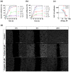
Effects of Pd2Spm and cisplatin on migration of MDA-MB-231 cells using scratch/wound healing assay. (a) Dose-dependent effect of Pd2Spm on migration of MDA-MB-231 cells expressed as wound confluence for tested Pd2Spm concentrations. (b) Dose-dependent effect of cisplatin on migration of MDA-MB-231 cells expressed as wound confluence for tested cisplatin concentrations. (c) Dose–response curve obtained from measuring the area under confluence curve (AUC) between 0 and 18 h of exposure to Pd2Spm or cisplatin. (d) Representative images from 5 independent experiments showing effects of 0 µM (control) and 50 µM of Pd2Spm or cisplatin on cell migration at 0, 3 and 24 h. Scale bar = 1000 µm. Data are expressed as mean ± SEM, n = 5. Data points with no visible error bars have errors smaller than the size of the symbol.
Figure 3
Figure 3
Pd2Spm suppresses tumor growth of MDA-MB-231 breast cancer xenografts. Graph of the effect of Pd2Spm treatment (5 mg/kg/day), cisplatin treatment (2 mg/kg/day) or vehicle (PBS + 0.5% DMSO) on MDA-MB-231 tumor volume. Drugs were administered intraperitoneally for 5 consecutive days in CBA nude female mice bearing subcutaneously implanted MDA-MB-231 triple-negative human breast cancer tumors. Data are expressed as mean tumor volume (mm3) ± SEM and analyzed with two-way ANOVA followed by Tukey’s multiple comparison test. * p < 0.05 Pd2Spm versus vehicle; *** p < 0.001 cisplatin versus vehicle.
Figure 4
Figure 4
(a) Representative micrographs of MDA-MB-231 breast cancer tumors stained with hematoxylin–eosin (H&E, black arrows show mitotic figures), Ki-67 nuclear staining and TUNEL assay staining (white arrows show TUNEL positive cells). (b) Quantitative analysis of mitotic figures. (c) Quantitative analysis of Ki-67 proliferative index. (d) Quantitative analysis of TUNEL-positive cells. Data are shown as box plots with medians analyzed with Kruskal–Wallis test followed by Dunn’s multiple comparisons test. ** p < 0.01, *** p < 0.001. Five random fields per tumor with at least 600 cells/field were analyzed. Scale bar = 100 µm. Pd2Spm 5 mg/kg/day (n = 7), cisplatin 2 mg/kg/day (n = 7), vehicle (n = 5).
Figure 5
Figure 5
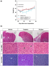
Impact of Pd2Spm, cisplatin or vehicle treatment on the body weight of MDA-MB-231 breast tumor-bearing mice. (a) Body weight changes of tumor-bearing mice treated intraperitoneally with either Pd2Spm (5 mg/kg/day), cisplatin (2 mg/kg/day) or vehicle (PBS + 0.5% DMSO) for 5 consecutive days. Data are expressed as mean weight (g) ± SEM. Two-way ANOVA followed by Tukey’s multiple comparisons test was used to analyze weight change in groups over the time, * p < 0.05 cisplatin versus vehicle. (b) Representative microphotographs of kidney and liver from animals treated with Pd2Spm, cisplatin or vehicle, showing absence of perirenal adipose tissues (white arrows) in cisplatin-treated animals. Hepatocytes of cisplatin-treated animals showing marked cytoplasmic vacuolization and hepatocellular glycogen deposits (black arrows). Scale bar (kidney) = 1000 µm. Scale bar (liver) = 100 µm.
Figure 6
Figure 6
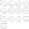
(a) Hematologic analysis and (b) Biochemical analysis of CBA nude female mice bearing subcutaneously implanted MDA-MB-231 triple-negative human breast cancer tumors treated with either Pd2Spm (5 mg/kg/day), cisplatin (2 mg/kg/day) or vehicle (PBS + 0.5% DMSO). Data are shown as individual values with medians, analyzed with Kruskal–Wallis test followed by Dunn’s multiple comparisons test. * p < 0.05, ** p < 0.01, ns = not significant. Pd2Spm (n = 7), cisplatin (n = 5), vehicle (n = 5).
Figure 7
Figure 7
Quantitative results of CAM angiogenesis in the presence of increasing concentrations (2–8 µM) of either cisplatin or Pd2Spm. (a) Parameters of early steps of angiogenesis: number of nodes, junctions and segments. (b) Parameters of later steps of angiogenesis: total length, branching length and segment length of vessels. The results are expressed as means of the fraction of the control ± SEM from 2 independent experiments (n = 2, 8 replicates). Significant differences from the vehicle control: * p < 0.05, ** p < 0.01, *** p < 0.001 (one-way ANOVA followed by Dunnett’s multiple comparison test).
Figure 8
Figure 8
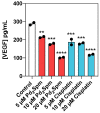
Quantitative results of VEGF secretion by MDA-MB-231 incubated for 12 h with Pd2Spm (red) or cisplatin (blue). Data are shown as means ± SEM (n = 2) and analyzed with one-way ANOVA followed by Dunnett’s multiple comparison test. Significant differences from the control: ** p < 0.01, *** p < 0.001, **** p < 0.0001.

References

    1. Sung H., Ferlay J., Siegel R.L., Laversanne M., Soerjomataram I., Jemal A., Bray F. Global Cancer Statistics 2020: GLOBOCAN Estimates of Incidence and Mortality Worldwide for 36 Cancers in 185 Countries. CA Cancer J. Clin. 2021;71:209–249. doi: 10.3322/caac.21660. - DOI - PubMed
    1. Wu Q., Siddharth S., Sharma D. Triple Negative Breast Cancer: A Mountain Yet to Be Scaled Despite the Triumphs. Cancers. 2021;13:3697. doi: 10.3390/cancers13153697. - DOI - PMC - PubMed
    1. Bianchini G., De Angelis C., Licata L., Gianni L. Treatment Landscape of Triple-Negative Breast Cancer—Expanded Options, Evolving Needs. Nat. Rev. Clin. Oncol. 2021:274. doi: 10.1038/s41571-021-00565-2. - DOI - PubMed
    1. Manjunath M., Choudhary B. Triple-negative Breast Cancer: A Run-through of Features, Classification and Current Therapies (Review) Oncol. Lett. 2021;22:512. doi: 10.3892/ol.2021.12773. - DOI - PMC - PubMed
    1. Yang R., Shi Y.-Y., Han X.-H., Liu S. The Impact of Platinum-Containing Chemotherapies in Advanced Triple-Negative Breast Cancer: Meta-Analytical Approach to Evaluating Its Efficacy and Safety. Oncol. Res. Treat. 2021;44:333–343. doi: 10.1159/000515353. - DOI - PubMed

LinkOut - more resources