. 2022 Jan 21;10(2):227.
doi: 10.3390/biomedicines10020227.

Antibacterial and Immunomodulatory Properties of Acellular Wharton's Jelly Matrix

Affiliations

Antibacterial and Immunomodulatory Properties of Acellular Wharton's Jelly Matrix

Marie Dubus et al. Biomedicines. .

Abstract

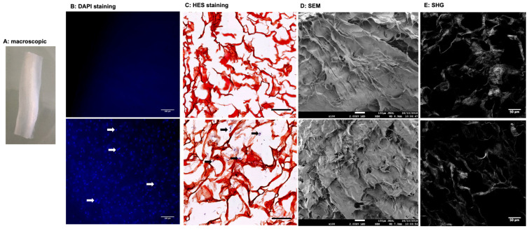
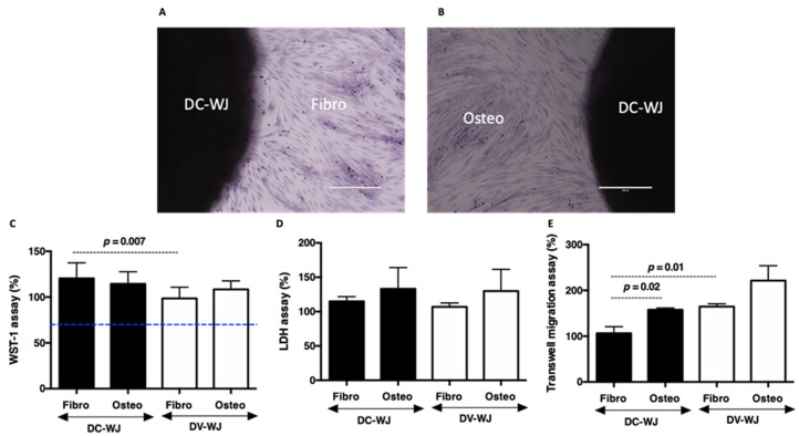
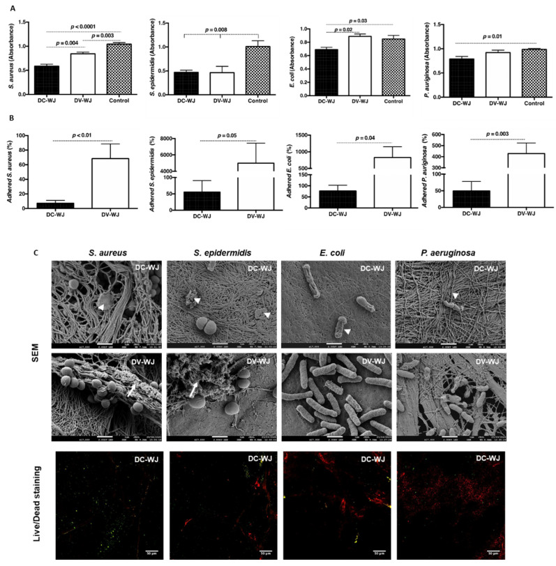
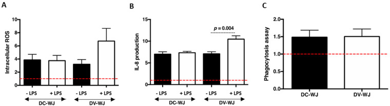
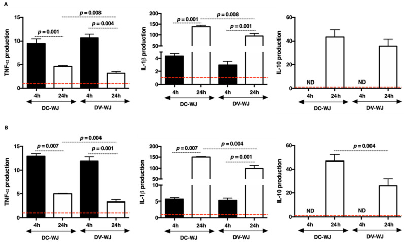
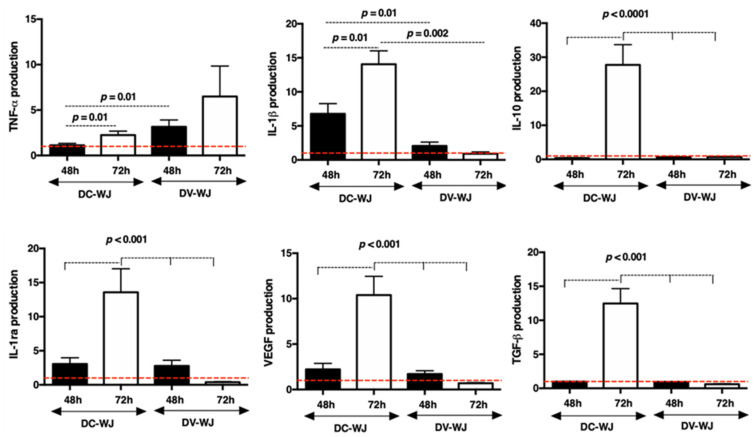
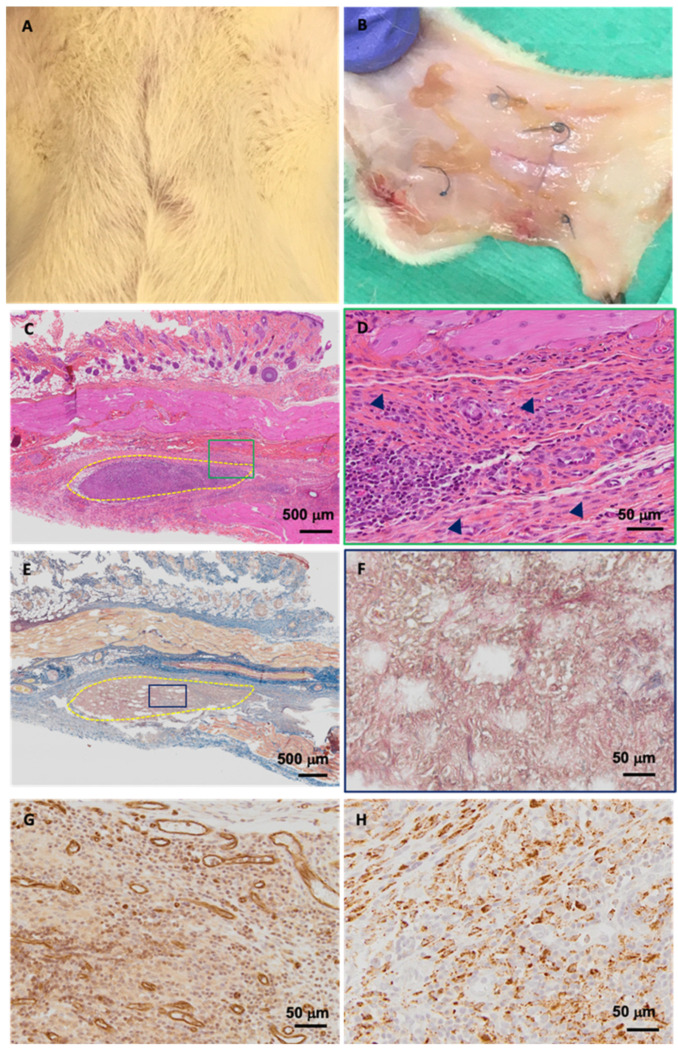
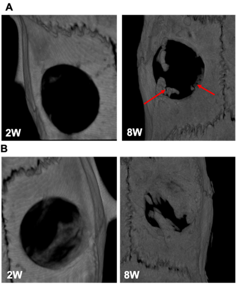
Of all biologic matrices, decellularized tissues have emerged as a promising tool in the field of regenerative medicine. Few empirical clinical studies have shown that Wharton's jelly (WJ) of the human umbilical cord promotes wound closure and reduces wound-related infections. In this scope, we herein investigated whether decellularized (DC)-WJ could be used as an engineered biomaterial. In comparison with devitalized (DV)-WJ, our results showed an inherent effect of DC-WJ on Gram positive (S. aureus and S. epidermidis) and Gram negative (E. coli and P. aeruginosa) growth and adhesion. Although DC-WJ activated the neutrophils and monocytes in a comparable magnitude to DV-WJ, macrophages modulated their phenotypes and polarization states from the resting M0 phenotype to the hybrid M1/M2 phenotype in the presence of DC-WJ. M1 phenotype was predominant in the presence of DV-WJ. Finally, the subcutaneous implantation of DC-WJ showed total resorption after three weeks of implantation without any sign of foreign body reaction. These significant data shed light on the potential regenerative application of DC-WJ in providing a suitable biomaterial for tissue regenerative medicine and an ideal strategy to prevent wound-associated infections.

Keywords: Wharton’s jelly; antibacterial; bioactivity; decellularization; immunomodulation.


Conflict of interest statement

The authors declare no conflict of interest.

Figures

Figure 1
Figure 1
Structural features of decellularized (upper line) and devitalized (lower line) Wharton’s Jelly matrix. (A): Macroscopical view of DC-WJ. (B): Fluorescence microscopy visualization of DAPI stained nuclei (scale bar = 100 µm), (C): Hematoxylin-eosin-Safran (HES) staining of paraffin-embedded sections. (scale bar = 200 µm). White and black arrows highlight stained nuclei (blue and purple color, respectively). (D): Scanning electron microscopy (SEM) views (scale bar = 100 µm) and (E): Two-photon excitation laser scanning confocal microscopy and second harmonic generation (SHG) (scale bar = 50 µm).
Figure 2
Figure 2
Biocompatibility of decellularized Wharton’s Jelly. (A,B): Pictures showing respectively crystal violet labelled fibroblasts (Fibro) and osteoblast (Osteo) growing in the presence of DC-WJ (scale bars = 400 µm). (C,D): Histograms reflecting percentage of cell viability and LDH release by damaged cells, respectively. Blue line indicates the threshold considered as an indicator of cytotoxic phenomenon, according to ISO standard (ISO/EN 10,993 part 5 guidelines), highlighting the absence of cytotoxic agents in both samples. (E): Transwell chemotaxis assay, showing a significant increase in fibroblast migration in the presence of DV-WJ compared DC-WJ (n = 6, Wilcoxon-Mann-Whitney test).
Figure 3
Figure 3
Antibacterial properties of decellularized and devitalized Wharton’s Jelly. (A): planktonic growth of bacteria in the presence of DC-WJ and DV-WJ. (B): Adhered bacteria on DC-WJ and DV-WJ. (n = 9, Wilcoxon-Mann-Whitney test). (C): Scanning electron microscopy pictures of bacteria adhered on DC-WJ (upper line) and on DV-WJ (middle line). Head arrows indicate damaged bacteria and full arrows indicate biofilm-like matrix (scale bars = 1 µm). Confocal observations of adhered bacteria on DC-WJ (lower line), in red dead bacteria and in green alive bacteria (scale bars = 50 µm).
Figure 4
Figure 4
Innate neutrophil responses to decellularized and devitalized Wharton’s Jelly. (A,B): Intracellular accumulation of ROS in neutrophils and IL-8 production by neutrophils in the presence of DC-WJ and DV-WJ. (C): Median fluorescence intensity of phagocyted Bioparticles. All the results were normalized to the basal production of neutrophils indicated by red dashed lines; n = 4, Wilcoxon-Mann-Whitney test.
Figure 5
Figure 5
Innate monocyte responses to decellularized and devitalized Wharton’s Jelly. (A): cytokine production following contact with DC-WJ and DV-WJ. (B): cytokine production following contact with DC-WJ and DV-WJ co-stimulated with LPS. Results were normalized to the basal monocyte production indicated by the red dashed lines; n = 6, Wilcoxon–Mann–Whitney test.
Figure 6
Figure 6
Macrophage polarization in the presence of decellularized and devitalized Wharton’s Jelly. Cytokine production following contact with DC- and DV-WJ. Results were normalized to DNA and M0 production, indicated by the red dashed lines; n = 4, Wilcoxon-Mann-Whitney test.
Figure 7
Figure 7
Subcutaneous biocompatibility of decellularized Wharton’s Jelly. (A,B): Macroscopical examination of the wound recovery and explanted tissue, respectively. (C,D): Hematoxylin-eosin-Safran (HES) staining of paraffin-embedded explant (delimited by dashed yellow line) at lower and higher magnifications, respectively. Head arrow point fibroblasts. (E,F): Masson trichrome staining of paraffin-embedded explant at lower and higher magnifications, respectively. Higher magnifications correspond to green and blue rectangles. (G): CD31 immunohistochemical staining and (H): CD68 immunohistochemical staining of the granulation tissue. (scale bar = 500 and 50 µm).
Figure 8
Figure 8
Calvaria bone defect regeneration. (A,B): MicroCT results following implantation of DC-WJ and DV-WJ, respectively. The two weeks (2 W) scans were obtained in vivo (35.8 µm) while eight weeks (8 W) scans were obtained ex vivo (17.9 µm). Red arrows indicate marginal bone regeneration in the presence of DC-WJ.

Similar articles

Cited by

References

    1. Edgar L., Pu T., Porter B., Aziz J.M., La Pointe C., Asthana A., Orlando G. Regenerative medicine, organ bioengineering and trans-plantation. J. Br. Surg. 2020;107:793–800. doi: 10.1002/bjs.11686. - DOI - PubMed
    1. Richbourg N.R., Peppas N.A., Sikavitsas V.I. Tuning the biomimetic behavior of scaffolds for regenerative medicine through surface modifications. J. Tissue Eng. Regen. Med. 2019;13:1275–1293. doi: 10.1002/term.2859. - DOI - PMC - PubMed
    1. Chan W.-I., Zhang G., Li X., Leung C.-H., Ma D.-L., Dong L., Wang C. Carrageenan activates monocytes via type-specific binding with interleukin-8: An implication for design of immuno-active biomaterials. Biomater. Sci. 2017;5:403–407. doi: 10.1039/C6BM00799F. - DOI - PubMed
    1. Yin J., Luan S. Opportunities and challenges for the development of polymer-based biomaterials and medical devices. Regen. Biomater. 2016;3:129–135. doi: 10.1093/rb/rbw008. - DOI - PMC - PubMed
    1. Londono R., Badylak S.F. Biologic Scaffolds for Regenerative Medicine: Mechanisms of In vivo Remodeling. Ann. Biomed. Eng. 2015;43:577–592. doi: 10.1007/s10439-014-1103-8. - DOI - PubMed

LinkOut - more resources