Review
. 2022 Feb 1;10(2):341.
doi: 10.3390/biomedicines10020341.

Mitochondrial Metabolism, Redox, and Calcium Homeostasis in Pulmonary Arterial Hypertension

Affiliations
Review

Mitochondrial Metabolism, Redox, and Calcium Homeostasis in Pulmonary Arterial Hypertension

Shuxin Liang et al. Biomedicines. .

Abstract

Pulmonary arterial hypertension (PAH) is a progressive disease characterized by elevated pulmonary arterial pressure due to increased pulmonary vascular resistance, secondary to sustained pulmonary vasoconstriction and excessive obliterative pulmonary vascular remodeling. Work over the last decade has led to the identification of a critical role for metabolic reprogramming in the PAH pathogenesis. It is becoming clear that in addition to its role in ATP generation, the mitochondrion is an important organelle that regulates complex and integrative metabolic- and signal transduction pathways. This review focuses on mitochondrial metabolism alterations that occur in deranged pulmonary vessels and the right ventricle, including abnormalities in glycolysis and glucose oxidation, fatty acid oxidation, glutaminolysis, redox homeostasis, as well as iron and calcium metabolism. Further understanding of these mitochondrial metabolic mechanisms could provide viable therapeutic approaches for PAH patients.

Keywords: metabolism; mitochondria; pulmonary hypertension.


Conflict of interest statement

The authors declare no conflict of interest.

Figures

Figure 1
Figure 1
Alterations of mitochondrial metabolic pathways and therapeutic targets for PAH treatment. Under normal conditions, glucose is converted to pyruvate via glycolysis. Pyruvate enters the mitochondria, where it is oxidized in the TCA cycle to generate ATP. In PAH, pyruvate is utilized for lactate production. The production of lactate in the presence of oxygen is known as “aerobic glycolysis” or the Warburg effect. In aerobic glycolysis, excess glucose uptake is diverted through PPP. Glutamine is another fuel source, which enters the mitochondria to replenish TCA intermediates and mobilize cellular energy, carbon, and nitrogen. FAs are the main energy source in the healthy adult heart. FA synthesis is started with the formation of malonyl-CoA by carboxylation of acetyl-CoA. Increased FAO inhibits glucose oxidation. HIF activates the transcription of genes encoding metabolic enzymes that mediate the glycolytic pathway. Red indicates increase, and blue indicates reduction. Abbreviations: ACACA, acetyl-CoA carboxylase; ACAT, acetyl-CoA acetyltransferase; Acetyl-CoA, acetyl coenzyme A; ACSL1, fatty acetyl-CoA L1; Acyl-CoA, acyl-coenzyme A; α-KG, α-ketoglutarate; CPT1, carnitine palmitoyltransferase 1; ENOL, enolase; Fructose-6P, fructose 6-phosphate; Glucose-6P, glucose-6-phosphate; G6PD, glucose-6-phosphate dehydrogenase; GLUT, glucose transporter; HIF, hypoxia-inducible factor; HK, hexokinase; LDHA, lactate dehydrogenase A; MCD, malonyl-CoA decarboxylase; MPC, mitochondrial pyruvate carrier; NADPH, reduced nicotinamide adenine dinucleotide phosphate; OAA, oxaloacetate; 6-P-gluconolactone, 6-phosphate-gluconolactone; PAH, pulmonary arterial hypertension; PDK, pyruvate dehydrogenase kinase; PEP, phosphoenolpyruvate; PFKFB, 6-phosphofructo-2-kinase/fructose-2,6-biphosphatase; PKM, pyruvate kinase M; PPP: pentose phosphate pathway; SLC1A5, solute carrier family 1 member 5; SLC7A5, solute carrier family 7 member 5; SucCoA, succinyl-coenzyme A; TCA, tricarboxylic acid.
Figure 2
Figure 2
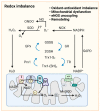
Redox signaling in pulmonary vasculature. GSH and Trx1-(SH)2 are oxidized to GSSG and Trx-S2, recycled in the redox cycle via the NADPH-dependent enzyme GR or TR, while NADP+ can be reduced to NADPH by G6PD in the cytoplasm. Once O2•− is formed by cytoplasmic NOXs and mitochondrial ETC, cytoplasmic SOD and mitochondrial MnSOD catalyze its dismutation into H2O2. GPx uses GSH to further reduce H2O2 to H2O. NO reacts with O2•− to form ONOO. In mitochondria, ~0.1~0.2% of the total oxygen accepts electrons from ETC to form O2•−. H2O2 is formed by the conversion of O2•− catalyzed by MnSOD or spontaneous dismutation. The production of excessive superoxide free radicals can lead to redox imbalance, mitochondrial damage, uncoupling of eNOS, and ultimately to impaired PAdiastolic function and remodeling. Abbreviations: ADP, adenosine diphosphate; ATP, adenosine triphosphate; eNOS, endothelial NO synthase; ETC, electron transport chain; G6PD, glucose-6-phosphate dehydrogenase; GPx, glutathione peroxidase; GR, glutathione reductase; GSH, reduced glutathione; GSSG, oxidized glutathione; H2O2, hydrogen peroxide; MnSOD, manganese superoxide dismutase; NAD(P)+/NAD(P)H, nicotinamide adenine dinucleotide (phosphate); NO, nitric oxide; NOX, NADPH oxidase; O2•−, superoxide anion; Prx1, peroxiredoxin 1; ONOO, peroxynitrite; ROS, reactive oxygen species; SOD, superoxide dismutase; TR, thioredoxin reductase; Trx-S2, oxidized thioredoxin; Trx-(SH)2, reduced thioredoxin.
Figure 3
Figure 3
Mitochondrial iron and calcium imbalance in PH. (A) Iron uptake and distribution are regulated by IRP, FPN1, and TfR1. In hypoxic and iron-deficient contexts, IRP1 is reduced to stabilize HIF-2α in vascular cells and subsequently induces the expression of ET-1, which regulates pulmonary vascular contraction and the proliferation of SMCs, cardiomyocytes, and fibroblasts. (B) Ca2+ transfer between ER and mitochondria is mediated by a multiprotein complex composed of IP3R1 in ER or RYR in SR, GRP75, and VDAC1 in OMM, and MUC in IMM. Mitochondrial Ca2+ uptake affects mitochondrial metabolism and promotes glucose oxidation via stimulating the Krebs cycle. The loss of MCU in PAH decreases the mitochondrial Ca2+ and simultaneously increases cytosolic Ca2+, inhibiting PDH and promoting a shift to glycolysis. ER: endoplasmic reticulum, ET-1: endothelin-1; FPN1: ferroportin 1, GLUT: glucose transporter, GRP75: 75 kDa glucose-regulated protein, HIF-2α: hypoxia-inducible factor 2α, IMM: inner mitochondrial membrane, IP3R1: inositol 1,4,5-trisphosphate receptor type 1, IRP1/2: iron regulatory protein 1/2, ISC: iron-sulfur cluster; ISPs: ISC-containing proteins, LIP: labile iron pool, MCU: mitochondrial calcium uniporter, OMM: outer mitochondrial membrane, RYR: ryanodine receptor, SERCA: sarco and endoplasmic reticulum calcium transporting ATPase, SR: sarcoplasmic reticulum, TCA: tricarboxylic acid, Tf: transferrin, TfR1: transferrin receptor 1, VDAC1: voltage-dependent anion-selective channel protein 1.

References

    1. Simonneau G., Montani D., Celermajer D.S., Denton C.P., Gatzoulis M.A., Krowka M., Williams P.G., Souza R. Haemodynamic definitions and updated clinical classification of pulmonary hypertension. Eur. Respir. J. 2019;53:1801913. doi: 10.1183/13993003.01913-2018. - DOI - PMC - PubMed
    1. Chan S.Y., Loscalzo J. Pathogenic mechanisms of pulmonary arterial hypertension. J. Mol. Cell Cardiol. 2008;44:14–30. doi: 10.1016/j.yjmcc.2007.09.006. - DOI - PMC - PubMed
    1. Tuder R.M., Archer S.L., Dorfmüller P., Erzurum S.C., Guignabert C., Michelakis E., Rabinovitch M., Schermuly R., Stenmark K.R., Morrell N.W. Relevant issues in the pathology and pathobiology of pulmonary hypertension. J. Am. Coll. Cardiol. 2013;62((Suppl. S25)):D4–D12. doi: 10.1016/j.jacc.2013.10.025. - DOI - PMC - PubMed
    1. Kierans S.J., Taylor C.T. Regulation of glycolysis by the hypoxia-inducible factor (HIF): Implications for cellular physiology. J. Physiol. 2021;599:23–37. doi: 10.1113/JP280572. - DOI - PubMed
    1. Tang H., Babicheva A., Mcdermott K.M., Gu Y., Ayon R.J., Song S., Wang Z., Gupta A., Zhou T., Sun X., et al. Endothelial HIF-2α contributes to severe pulmonary hypertension due to endothelial-to-mesenchymal transition. Am. J. Physiol. Lung Cell. Mol. Physiol. 2018;314:L256–L275. - PMC - PubMed

LinkOut - more resources