Review
. 2022 Feb 1;10(2):347.
doi: 10.3390/biomedicines10020347.

Pathophysiology of Perinatal Asphyxia in Humans and Animal Models

Affiliations
Review

Pathophysiology of Perinatal Asphyxia in Humans and Animal Models

Daniel Mota-Rojas et al. Biomedicines. .

Abstract

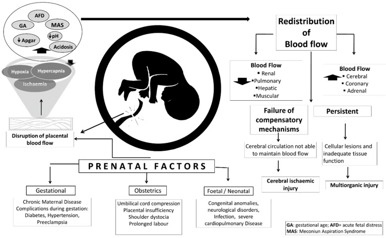
Perinatal asphyxia is caused by lack of oxygen delivery (hypoxia) to end organs due to an hypoxemic or ischemic insult occurring in temporal proximity to labor (peripartum) or delivery (intrapartum). Hypoxic-ischemic encephalopathy is the clinical manifestation of hypoxic injury to the brain and is usually graded as mild, moderate, or severe. The search for useful biomarkers to precisely predict the severity of lesions in perinatal asphyxia and hypoxic-ischemic encephalopathy (HIE) is a field of increasing interest. As pathophysiology is not fully comprehended, the gold standard for treatment remains an active area of research. Hypothermia has proven to be an effective neuroprotective strategy and has been implemented in clinical routine. Current studies are exploring various add-on therapies, including erythropoietin, xenon, topiramate, melatonin, and stem cells. This review aims to perform an updated integration of the pathophysiological processes after perinatal asphyxia in humans and animal models to allow us to answer some questions and provide an interim update on progress in this field.

Keywords: brain injury; human and animal models; hypoxic–ischemic encephalopathy; meconium aspiration syndrome; perinatal asphyxia.


Conflict of interest statement

All the authors declare that there is no conflict of interest or ethical concern in this article.

Figures

Figure 1
Figure 1
Prenatal risk factors and effect on cerebral ischemic injury.
Figure 2
Figure 2
Fetal asphyxia in human and non-human animals, organ injury and physiological imbalances in response to hypoxia–ischemia. Image (A) summarizes the neuronal damage at a biochemical and pathological level. Image (B) shows damage in other vital organs. In image (C), the physiological imbalances are appreciated due to the drastic reduction of oxygen to the fetus, and promote the expulsion of meconium. In letter (D) some adverse effects if the newborn survives.
Figure 3
Figure 3
Neonatal piglets with perinatal hypoxia syndrome. When the neonate goes through a severe hypoxia process, it displays bradycardia, yellow to greenish meconium staining, tachypnea, lactic acidemia, hypothermia, hypoglycemia, adynamia, and flaccid muscle tone. Neonates with this perinatal syndrome do not recover, since they do not connect with the teat and are sluggish, which is why they die in the following postpartum hours. There is no neonatal intensive therapy area on pig farms.
Figure 4
Figure 4
Periventricular white matter greyish discoloration and edema in a male, 32 weeks of gestational age old, with HIE. (Courtesy: María de Lourdes Cabrera-Muñoz, MD, Department of Pathology, Hospital Infantil de México Federico Gómez).
Figure 5
Figure 5
(A). Bilateral basal ganglia necrosis in a male at 39.6 weeks gestation with severe perinatal asphyxia secondary to congenital heart disease. (B). Neuronal necrosis and calcification (arrows). (C) Withe matter infarct (*) HE 40X. (Courtesy: María de Lourdes Cabrera-Muñoz MD. Department of Pathology, Hospital Infantil de México Federico Gómez).
Figure 6
Figure 6
(a) High cerebral oxygenation on NIRS * with (b) a low-electrical-activity aEEG background ** in severe HIE neonates on hypothermia treatment at 12 h of age has a 91%positive predictive value for long-term adverse neurological outcome (magnetic resonance imaging and neurodevelopmental assessment at 18 months of age), and the absence of these results in a negative predictive value of 100%. NIRS: near infrared spectroscopy; aEEG: amplitude-integrated electroencephalography; rScO2: regional cerebral oxygenation; cFTOE: cerebral fractional tissue oxygen extraction. Courtesy: Daniel Ibarra-Ríos, MD, Department of Neonatology, Hospital Infantil de México Federico Gómez.
Figure 7
Figure 7
Magnetic resonance: (a) axial image of sequence enhanced in T1 towards convexity and (b) enhanced in FLAIR, where hyperintensity of the bilateral semioval centers is observed, as well as a decrease in volume in bordering territory in parietal regions and in the smaller frontal portion with retraction of the lateral ventricles associated with hyperintensity of the periventricular white matter. Courtesy: Eduardo M. Flores Armas, MD, Department of Medical Imaging. Cranial ultrasound: (c) low RI (<0.55) in normothermic infants or after rewarming has an 84% positive predictive value for death or disability. Courtesy: Daniel Ibarra-Ríos, MD, Department of Neonatology, Hospital Infantil de México Federico Gómez. ACA: anterior cerebral artery; MCA: medial cerebral artery; RI: resistive index.
Figure 8
Figure 8
Male of 34 weeks of gestational with multiorgan damage (heart, lungs, liver, gut, and kidneys) after severe perinatal asphyxia. (Courtesy: Dina Villanueva-García, MD, Department of Neonatology, Hospital Infantil de México Federico Gómez).
Figure 9
Figure 9
Thermograms of newborn piglets with severe hypoxia and meconium staining on the skin to a severe degree. It is important to dry the newborn immediately, since the humidity of the amniotic fluid favors a rapid drop in body temperature. The areas marked in yellow on the thermograms indicate temperatures between 28 and 32 °C, especially in images (AC), where a marked drop in temperature is seen in peripheral areas of the auricular pavilion, thoracic limbs, and especially the face, particularly the snout. In thermographic image (D), we can see a piglet stained with severe meconium grade, with umbilical-cord rupture, a failing vitality score, and severe hypothermia, despite having been dried. It is important to note that different temperatures are seen depending on the body region. In the frontal area of the head and left pectoral region, the highest surface temperatures are observed (35.5 °C). In yellow (image (D)), the proximal region of the snout and the auricular pavilion (28–29 °C) are distinguished, and in the distal region of the thoracic limbs and the distal snout area (in blue), the lowest temperature ranges of 18–19 °C are shown. The use of infrared thermography is essential to understanding the changes in the vascular microcirculation in the study of hypothermia in newborns with asphyxia.

References

    1. WHO Newborns: Reducing Mortality. [(accessed on 11 November 2021)]. Available online: https://www.who.int/news-room/fact-sheets/detail/newborns-reducing-morta....
    1. Bhutta Z.A., Das J.K., Bahl R., Lawn J.E., Salam R.A., Paul V.K., Sankar M.J., Blencowe H., Rizvi A., Chou V.B., et al. Can available interventions end preventable deaths in mothers, newborn babies, and stillbirths, and at what cost? Lancet. 2014;384:347–370. doi: 10.1016/S0140-6736(14)60792-3. - DOI - PubMed
    1. Lawn J.E., Cousens S., Zupan J., Lancet Neonatal Survival Steering Team 4 million neonatal deaths: When? Where? Why? Lancet. 2005;365:891–900. doi: 10.1016/S0140-6736(05)71048-5. - DOI - PubMed
    1. Liu L., Johnson H.L., Cousens S., Perin J., Scott S., Lawn J.E., Rudan I., Campbell H., Cibulskis R., Li M., et al. Global, regional, and national causes of child mortality: An updated systematic analysis for 2010 with time trends since 2000. Lancet. 2012;379:2151–2161. doi: 10.1016/S0140-6736(12)60560-1. - DOI - PubMed
    1. Perez A., Ritter S., Brotschi B., Werner H., Caflisch J., Martin E., Latal B. Long-Term Neurodevelopmental Outcome with Hypoxic-Ischemic Encephalopathy. J. Pediatr. 2013;163:454–459.e1. doi: 10.1016/j.jpeds.2013.02.003. - DOI - PubMed