. 2022 Feb 8;10(2):403.
doi: 10.3390/biomedicines10020403.

Decellularized Dental Pulp, Extracellular Vesicles, and 5-Azacytidine: A New Tool for Endodontic Regeneration

Affiliations

Decellularized Dental Pulp, Extracellular Vesicles, and 5-Azacytidine: A New Tool for Endodontic Regeneration

Francesca Diomede et al. Biomedicines. .

Abstract

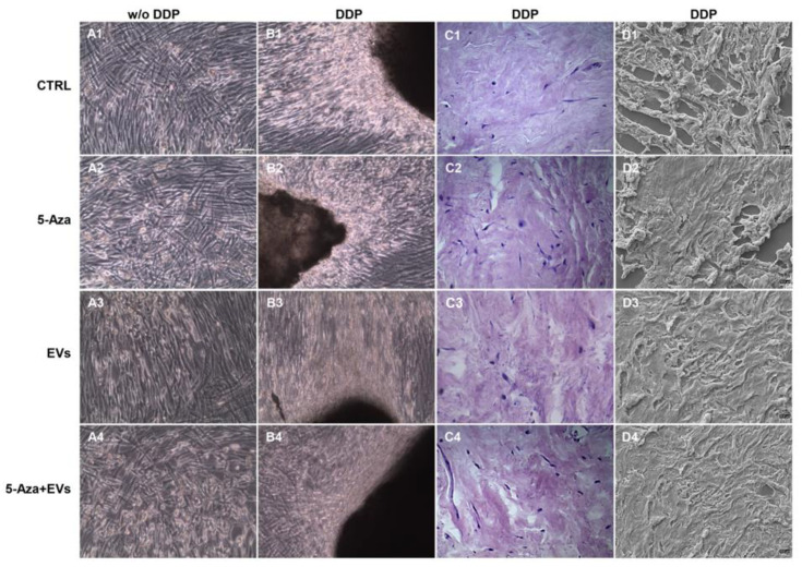
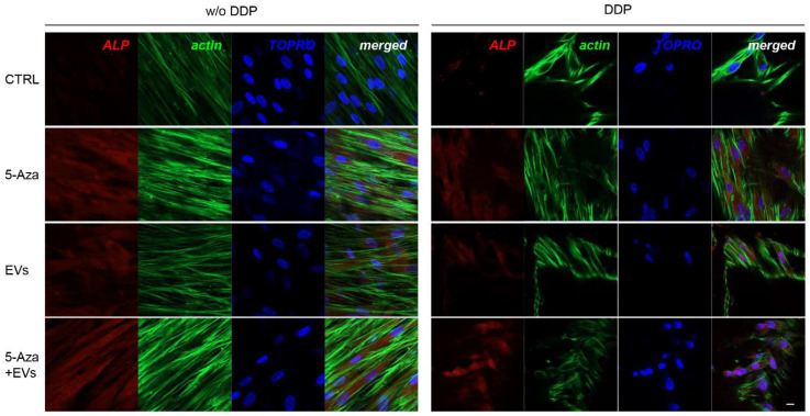
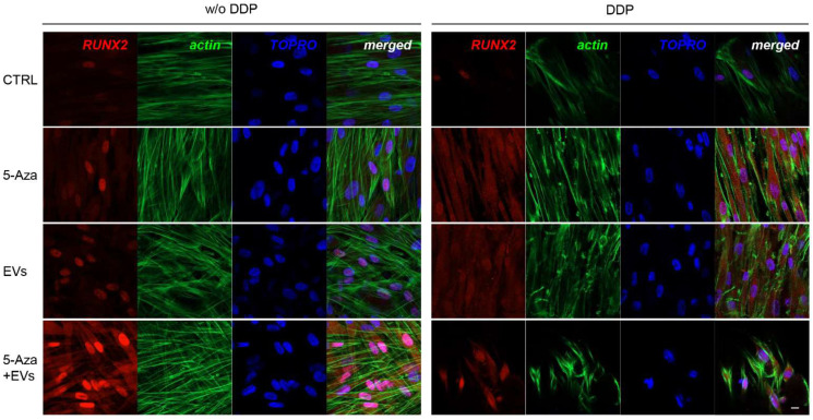
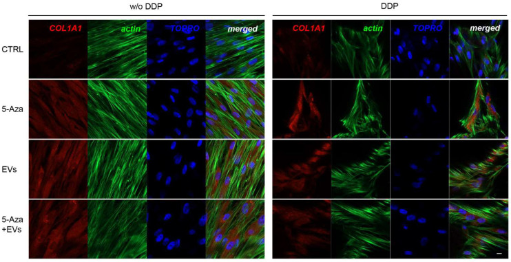
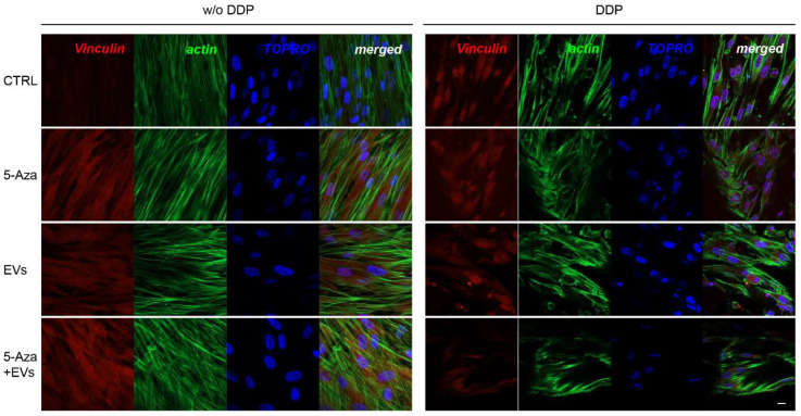
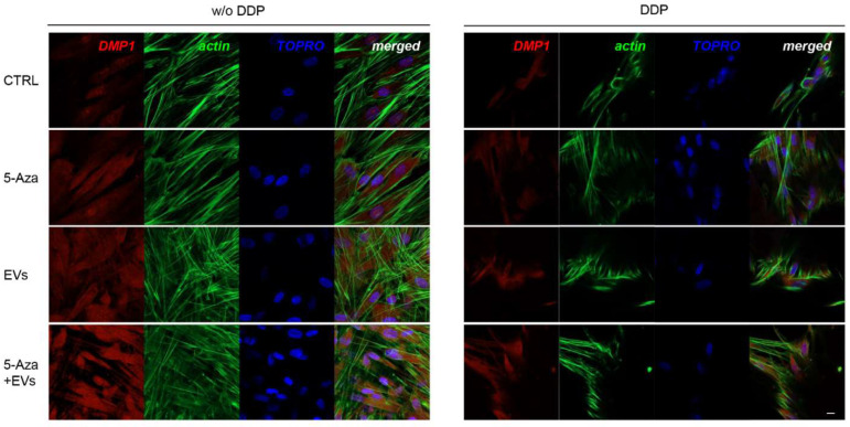
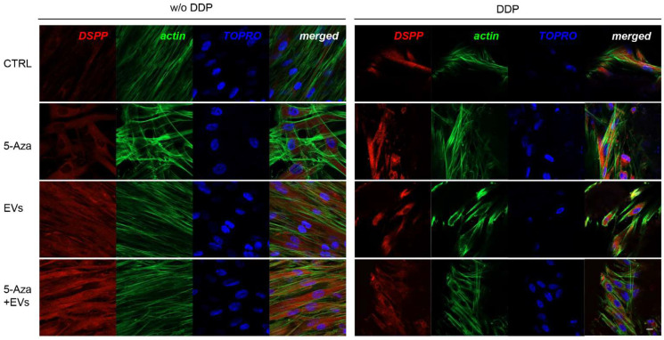
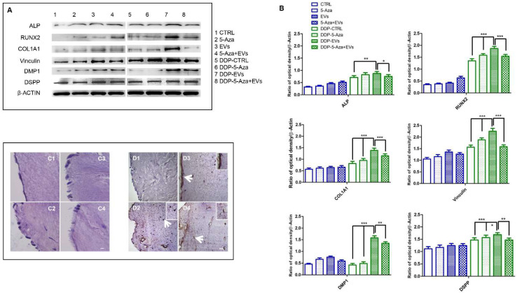
Dental pulp is a major component of the dental body that serves to maintain the tooth life and function. The aim of the present work was to develop a system that functions as a growth-permissive microenvironment for dental pulp regeneration using a decellularized dental pulp (DDP) matrix, 5-Aza-2'-deoxycytidine (5-Aza), and Extracellular Vesicles (EVs) derived from human Dental Pulp Stem Cells (hDPSCs). Human dental pulps extracted from healthy teeth, scheduled to be removed for orthodontic purpose, were decellularized and then recellularized with hDPSCs. The hDPSCs were seeded on DDP and maintained under different culture conditions: basal medium (CTRL), EVs, 5-Aza, and EVs+-5-Aza. Immunofluorescence staining and Western blot analyses were performed to evaluate the proteins' expression related to dentinogenesis, such as ALP, RUNX2, COL1A1, Vinculin, DMP1, and DSPP. Protein contents found in the DDP recellularized with hDPSCs were highly expressed in samples co-treated with EVs and 5-Aza compared to other culture conditions. This study developed a DDP matrix loaded by hDPSCs in co-treatment with EVs, which might enhance the dentinogenic differentiation with a high potentiality for endodontic regeneration.

Keywords: 5-azacytidine; decellularized dental pulp; endodontic regeneration; extracellular matrix scaffold; extracellular vesicles.


Conflict of interest statement

The authors declare no conflict of interest.

Figures

Figure 1
Figure 1
Morphological and cell distribution analyses in DDP and DP. (A,B) Images of toluidine blue staining of (A) DDP and (B) DP. Cell nuclei are clearly visible in DP sample when compared to DDP. Representative images are shown. (C,D) Images obtained at confocal laser scanning microscopy showed the presence of nuclei stained in blue in the DP sample, while in DDP the staining for cell nuclei was negative. Scale bar = 20 μm.
Figure 2
Figure 2
Morphological evaluation of hDPSCs cultured with and without DDP. (A1A4) Human DPSCs cultured without DDP observed under light microscopy in all conditions considered in the study. (A1) Cells cultured with basal medium (CTRL), (A2) cells cultured with 5-Aza (5-Aza), (A3) cells cultured with EVs (EVs), and (A4) cells cultured with EVs+5-Aza (EVs+5-Aza). (B1B4) Human DPSCs cultured on DDP observed under light microscopy in all conditions considered in the study. (B1) Cells cultured on DDP with basal medium (DDP-CTRL), (B2) cells cultured on DDP with 5-Aza (DDP-5-Aza), (B3) cells cultured on DDP and treated with EVs (DDP-EVs), and (B4) cells cultured on DDP with EVs+5-Aza (DDP-EVs+5-Aza). (C1C4) Haematoxylin-eosin staining of hDPSCs cultured on DDP in all conditions considered in the study. (C1) Cells cultured on DDP with basal medium (DDP-CTRL), (C2) cells cultured on DDP with 5-Aza (DDP-5-Aza), and (C3) cells cultured on DDP and treated with EVs (DDP-EVs), (C4) Cells cultured on DDP with EVs+5-Aza (DDP-EVs+5-Aza). (D1D4) Human DPSCs cultured on DDP observed via scanning electron microscopy in all conditions considered in the study. (D1) Cells cultured on DDP with basal medium (DDP-CTRL), (D2) cells cultured on DDP with 5-Aza (DDP-5-Aza), (D3) cells cultured on DDP and treated with EVs (DDP-EVs), and (D4) cells cultured on DDP with EVs+5-Aza (DDP-EVs+5-Aza). Scale bar: 100 µm.
Figure 3
Figure 3
Immunofluorescence analysis of ALP expression. Left panel shows the images of hDPSCs (CTRL), hDPSCs+5-Aza (5-Aza), hDPSCs+EVs (EVs), and hDPSCs+5-Aza+EVs (5-Aza+EVs) cultured without DDP. Right panel shows the images of hDPSCs (CTRL), hDPSCs+5-Aza (5-Aza), hDPSCs+EVs (EVs), and hDPSCs+5-Aza+EVs (5-Aza+EVs) cultured without DDP. Red fluorescence: ALP marker; green fluorescence: cytoskeleton actin; blue fluorescence: cell nuclei using TOPRO; merged image: overlap of all above mentioned channels. Representative images are shown. Scale bar: 20 µm.
Figure 4
Figure 4
Immunofluorescence analysis of RUNX2 expression. Left panel shows the images of hDPSCs (CTRL), hDPSCs+5-Aza (5-Aza), hDPSCs+EVs (EVs), and hDPSCs+5-Aza+EVs (5-Aza+EVs) cultured without DDP. Right panel shows the images of hDPSCs (CTRL), hDPSCs+5-Aza (5-Aza), hDPSCs+EVs (EVs), and hDPSCs+5-Aza+EVs (5-Aza+EVs) cultured without DDP. Red fluorescence: ALP marker; green fluorescence: cytoskeleton actin; blue fluorescence: cell nuclei using TOPRO; merged image: overlap of all above mentioned channels. Representative images are shown. Scale bar: 20 µm.
Figure 5
Figure 5
Immunofluorescence analysis of COL1A1 expression. Left panel showes the images of hDPSCs (CTRL), hDPSCs+5-Aza (5-Aza), hDPSCs+EVs (EVs), and hDPSCs+5-Aza+EVs (5-Aza+EVs) cultured without DDP. Right panel shows the images of hDPSCs (CTRL), hDPSCs+5-Aza (5-Aza), hDPSCs+EVs (EVs), and hDPSCs+5-Aza+EVs (5-Aza+EVs) cultured without DDP. Red fluorescence: ALP marker; green fluorescence: cytoskeleton actin; blue fluorescence: cell nuclei using TOPRO; merged image: overlap of all above mentioned channels. Representative images are shown. Scale bar: 20 µm.
Figure 6
Figure 6
Immunofluorescence analysis of Vinculin expression. Left panel shows the images of hDPSCs (CTRL), hDPSCs+5-Aza (5-Aza), hDPSCs+EVs (EVs), and hDPSCs+5-Aza+EVs (5-Aza+EVs) cultured without DDP. Right panel shows the images of hDPSCs (CTRL), hDPSCs+5-Aza (5-Aza), hDPSCs+EVs (EVs), and hDPSCs+5-Aza+EVs (5-Aza+EVs) cultured without DDP. Red fluorescence: ALP marker; green fluorescence: cytoskeleton actin; blue fluorescence: cell nuclei using TOPRO; merged image: overlap of all above mentioned channels. Representative images are shown. Scale bar: 20 µm.
Figure 7
Figure 7
Immunofluorescence analysis of DMP1 expression. Left panel shows the images of hDPSCs (CTRL), hDPSCs+5-Aza (5-Aza), hDPSCs+EVs (EVs), and hDPSCs+5-Aza+EVs (5-Aza+EVs) cultured without DDP. Right panel shows the images of hDPSCs (CTRL), hDPSCs+5-Aza (5-Aza), hDPSCs+EVs (EVs), and hDPSCs+5-Aza+EVs (5-Aza+EVs) cultured without DDP. Red fluorescence: ALP marker; green fluorescence: cytoskeleton actin; blue fluorescence: cell nuclei using TOPRO; merged image: overlap of all above mentioned channels. Representative images are shown. Scale bar: 20 µm.
Figure 8
Figure 8
Immunofluorescence analysis of DSPP expression. Left panel shows the images of hDPSCs (CTRL), hDPSCs+5-Aza (5-Aza), hDPSCs+EVs (EVs), and hDPSCs+5-Aza+EVs (5-Aza+EVs) cultured without DDP. Right panel shows the images of hDPSCs (CTRL), hDPSCs+5-Aza (5-Aza), hDPSCs+EVs (EVs), and hDPSCs+5-Aza+EVs (5-Aza+EVs) cultured without DDP. Red fluorescence: ALP marker; green fluorescence: cytoskeleton actin; blue fluorescence: cell nuclei using TOPRO; merged image: overlap of all above mentioned channels. Representative images are shown. Scale bar: 20 µm.
Figure 9
Figure 9
Odontoblast differentiation. (A) Specific Western blot bands of ALP, RUNX2, COL1A1, Vinculin, DMP1. and DSPP evaluated in all sample groups. (B) Densitometric analyses of ALP, RUNX2, COL1A1, Vinculin, DMP1, and DSPP. β-actin was used as loading control. Haematoxylin-eosin staining in all sample groups. Haematoxylin-eosin staining of (C1) hDPSCs cultured on DDP, (C2) hDPSCs cultured on DDP with 5-Aza, (C3) hDPSCs with EVs, and (C4) hDPSCs cultured on DDP with EVs+5-Aza. Immunoistochemical analysis of DSPP expression in (D1) hDPSCs cultured on DDP, (D2) hDPSCs cultured on DDP with 5-Aza, (D3) hDPSCs with EVs, and (D4) hDPSCs cultured on DDP with EVs+5-Aza. Arrows indicate the magnified inset area. Scale bar: 100 µm. Significant statistical differences: ALP DDP-EVs vs. DDP-CTRL ** p < 0.01, DDP-EVs vs. DDP-5-Aza+EVs * p < 0.05; RUNX2, COL1A1, Vinculin, DDP-EVs vs. DDP-CTRL *** p < 0.001, DDP-EVs vs. DDP-5-Aza *** p < 0.001, DDP-EVs vs. DDP-5-Aza+EVs *** p < 0.001; DMP1 DDP-EVs vs. DDP-CTRL *** p < 0.001, DDP-EVs vs. DDP-5-Aza *** p < 0.001, DDP-EVs vs. DDP-5-Aza+EVs ** p < 0.01; DSPP DDP-EVs vs. DDP-CTRL *** p < 0.001, DDP-EVs vs. DDP-5-Aza * p < 0.05, DDP-EVs vs. DDP-5-Aza+EVs ** p < 0.01.

References

    1. Petersen P.E., Bourgeois D., Ogawa H., Estupinan-Day S., Ndiaye C. The global burden of oral diseases and risks to oral health. Bull. World Health Organ. 2005;83:661–669. - PMC - PubMed
    1. Huang C.C., Narayanan R., Alapati S., Ravindran S. Exosomes as biomimetic tools for stem cell differentiation: Applications in dental pulp tissue regeneration. Biomaterials. 2016;111:103–115. doi: 10.1016/j.biomaterials.2016.09.029. - DOI - PMC - PubMed
    1. Ghasemi-Mobarakeh L., Beigi M.H., Ebrahimi-Kahrizsangi R., Karbalaie K., Nasr-Esfahani M.H. Dental Pulp Stem Cells and Nanofibrous Scaffolds for Application in Tissue Engineering. Int. J. Artif. Organs. 2011;34:686.
    1. Cordeiro M.M., Dong Z., Kaneko T., Zhang Z., Miyazawa M., Sbl S., Smitb A.J., Nor J.E. Dental pulp tissue engineering with stem cells from exfoliated deciduous teeth. J. Endod. 2008;34:962–969. doi: 10.1016/j.joen.2008.04.009. - DOI - PubMed
    1. Qiao L., Kong Y., Shi Y.S., Sun A.J., Ji R.J., Huang C., Li Y.Q., Yang X.Q. Synergistic effects of adipose-derived stem cells combined with decellularized myocardial matrix on the treatment of myocardial infarction in rats. Life Sci. 2019;239:116891. doi: 10.1016/j.lfs.2019.116891. - DOI - PubMed

LinkOut - more resources