. 2022 Jan 29;11(2):178.
doi: 10.3390/antibiotics11020178.

Repurposing α-Adrenoreceptor Blockers as Promising Anti-Virulence Agents in Gram-Negative Bacteria

Affiliations

Repurposing α-Adrenoreceptor Blockers as Promising Anti-Virulence Agents in Gram-Negative Bacteria

Ahmad J Almalki et al. Antibiotics (Basel). .

Abstract

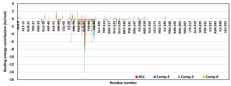
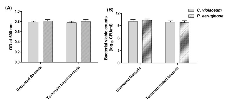
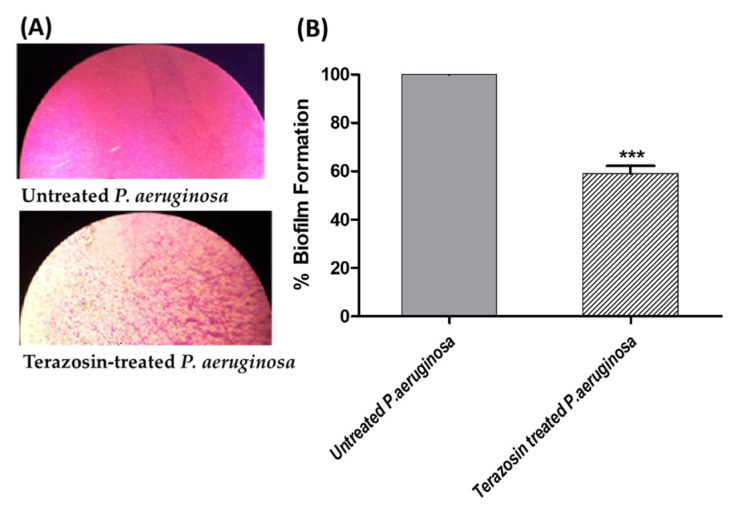
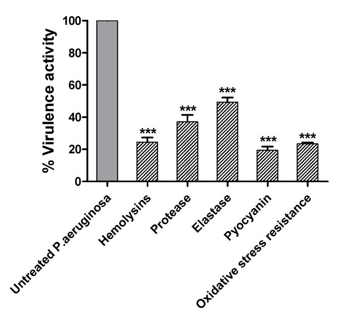
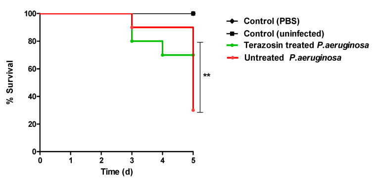
Antimicrobial resistance is among the world's most urgent public health problems. Diminishing of the virulence of bacteria is a promising approach to decrease the development of bacterial resistance. Quorum sensing (QS) systems orchestrate the bacterial virulence in inducer-receptors manner. Bacteria can spy on the cells of the host by sensing adrenergic hormones and other neurotransmitters, and in turn, these neurotransmitters can induce bacterial pathogenesis. In this direction, α-adrenergic blockers were proposed as an anti-virulence agents through inhibiting the bacterial espionage. The current study aimed to explore the α-blockers' anti-QS activities. Within comprehensive in silico investigation, the binding affinities of seven α-adrenoreceptor blockers were evaluated towards structurally different QS receptors. From the best docked α-blockers into QS receptors, terazosin was nominated to be subjected for further in vivo and in vitro anti-QS and anti-virulence activities against Chromobacterium violaceum and Pseudomonas aeruginosa. Terazosin showed a significant ability to diminish the QS-controlled pigment production in C. violaceum. Moreover, Terazosin decreased the P. aeruginosa biofilm formation and down-regulated its QS-encoding genes. Terazosin protected mice from the P. aeruginosa pathogenesis. In conclusion, α-adrenergic blockers are proposed as promising anti-virulence agents as they hinder QS receptors and inhibit bacterial espionage.

Keywords: Pseudomonas aeruginosa; bacterial virulence; quorum sensing; terazosin; α-adrenoreceptor blockers.


Conflict of interest statement

The authors declare no conflict of interest.

Figures

Figure 1
Figure 1
Molecular binding interaction of the ligand–protein complexes. (A) Cartoon and surface representation of P. aeruginosa QscR (PDB; 3SZT), showing an overlay of investigated α-adrenoreceptor hits (yellow lines) over reported reference inhibitor, HLC (magenta sticks), at the QS protein’s substrate binding site of protomer-B (green). The binding site involves the small (more polar) and larger (more hydrophobic) sub-pockets. At protomer-A (yellow), the O-C12-HSL co-crystalline ligand is represented as magenta spheres. (B) Predicted docking poses of the examined ligands (sticks). Residues within 5 Å radius of the in complex ligands were only displayed, colored based on their respective Qs subsite location, and sequentially labeled with numbers. For clarity, non-polar hydrogens are removed, while hydrogen bonding was represented as red dashed lines.
Figure 2
Figure 2
Molecular binding interaction of the ligand–protein complexes. (A) Cartoon and surface representation of C. violaceum CviR (PDB; 3QP5), showing an overlay of investigated α-adrenoreceptor hits (yellow lines) over reported reference inhibitor, HLC (magenta sticks), at the QS protein’s substrate binding site of protomer B (green). The binding site involves the small (more polar) and larger (more hydrophobic) sub-pockets. At protomer A (yellow), the co-crystalline ligand, HLC, is represented as magenta spheres. (B) Predicted docking poses of the investigated ligands (sticks). Residues within 5Å radius of the in complex ligands were only displayed, colored based on their respective Qs subsite location, and labeled with sequence number. For clarity, non-polar hydrogens were removed, while hydrogen bonding was depicted as red dashed lines.
Figure 3
Figure 3
Analysis of ligand–QscR P. aeruginosa model stability across the 200 ns explicit MD simulations. Alpha carbon RMSD (Å) trajectories of (A) P. aeruginosa QscR proteins; (B) α-adrenoreceptor and control inhibitors, against MD simulation time (ns). (C) Overlaid frames of the ligand–QscR P. aeruginosa protein complexes at initial (0 ns) and end (200 ns) of MD runs. The left, middle, and right panels are for HLC, Comp. 5, and Comp. 6 protein complexes, respectively. Both the ligands (sticks) and P. aeruginosa QscR proteins (cartoon) are illustrated in green or red, corresponding to the initial and end extracted frames, respectively.
Figure 4
Figure 4
Residue–wise global stability analysis of P. aeruginosa QscR proteins in relation to ΔRMSF trajectories across the entire 200 ns MD simulation timeframes. The estimated ΔRMSFs are illustrated as a function of the P. aeruginosa QscR protein residue numbers, where the latter are calculated assuming independent MD simulation of P. aeruginosa QscR apo/non-liganded states against the complexed holo proteins in bound to α-adrenoreceptor or reference inhibitors.
Figure 5
Figure 5
Residue–wise binding energy contributions within the total free–binding energy calculation of ligand–QscR P. aeruginosa complexes. Binding energy contributions are illustrated as a function of QscR P. aeruginosa protein residue numbers.
Figure 6
Figure 6
Analysis of ligand–CviR C. violaceum model stability across the 200 ns explicit MD simulations. Alpha carbon RMSD (Å) trajectories of (A) CviR C. violaceum proteins; (B) α-adrenoreceptor and control inhibitors, against MD simulation time (ns). (C) Overlaid frames of the ligand–CviR C. violaceum protein complexes at the start (0 ns) and end (200 ns) of MD runs. The left, middle, and right panels are for HLC, Comp. 4, Comp. 5, and Comp. 6 protein complexes, respectively. Both the ligands (sticks) and C. violaceum CviR proteins (cartoon) are illustrated in green or red, corresponding to the start and end of the extracted frames, respectively.
Figure 7
Figure 7
Residue–wise global stability analysis of C. violaceum CviR proteins in relation to ΔRMSF trajectories across the entire 200 ns MD simulation timeframes. The estimated ΔRMSFs are illustrated as a function of the C. violaceum CviR protein residue numbers, where the latter are calculated considering independent MD simulation of C. violaceum CviR apo/non-liganded states against the complexed/holo proteins in bound to α-adrenoreceptor or reference inhibitors.
Figure 8
Figure 8
Residue–wise binding energy contributions within the total free–binding energy calculation of ligand–CviR C. violaceum complexes. Binding energy contributions are illustrated as a function of CviR C. violaceum protein residue numbers.
Figure 9
Figure 9
Effect of terazosin on C. violaceum or P. aeruginosa growth. (A) The bacterial growth turbidities in the presence or absence of 1/4 MIC of terazosin were measured at OD 600 nm. (B) Viable count of terazosin-treated and control untreated bacterial cultures after overnight incubation. There was no significant effect of terazosin at sub-MIC on C. violaceum or P. aeruginosa growth.
Figure 10
Figure 10
Terazosin at sub-MIC effect on the production of QS-controlled violacein pigment. C. violaceum CV026 was allowed to grow in the absence or presence of terazosin at sub-MIC. The produced violacein was extracted by DMSO and the absorbances were evaluated at 590 nm. Terazosin significantly diminished the production of violacein (***: p ≤ 0.001).
Figure 11
Figure 11
Terazosin decreased the expression of virulence and QS genes of P. aeruginosa. RNA of P. aeruginosa, treated or not with terazosin at sub–MIC, was isolated, and the expression of each gene was normalized to the housekeeping gene rplU gene. The test was performed in triplicate and the results were expressed as mean ± standard error. The one-way ANOVA test, followed by Dunnett’s multiple comparison test, was used for statistical analysis. Terazosin significantly downregulated the expressions of all tested genes, ***: p ≤ 0.001.
Figure 12
Figure 12
Terazosin diminished the formation of biofilm in P. aeruginosa. A crystal violet method was used to stain the biofilm-forming cells in the absence or presence of terazosin at sub-MIC. (A) Light microscopic images showed a few dispersed adhered cells when treated with terazosin at sub-MIC. (B) The absorbance of the crystal violet staining the biofilm-forming cells in the absence or presence of terazosin at sub-MIC was measured at 590 nm. The results are presented as the percentage change from the untreated P. aeruginosa control. The Student’s t-test was employed to test the significance; terazosin significantly reduced biofilm formation, ***: p ≤ 0.001.
Figure 13
Figure 13
Terazosin diminished the (A) swarming and (B) swimming of P. aeruginosa motility. The swarming or swimming motility zones were measured in the absence or presence of terazosin at sub-MIC. The test was repeated in triplicate. The Student’s t-test was used to test the significance. Terazosin significantly diminished the P. aeruginosa motility, ***: p ≤ 0.001.
Figure 14
Figure 14
Terazosin decreased the P. aeruginosa virulence. Terazosin at sub-MIC significantly reduced the production of hemolysins, protease, and elastase enzymes, as well as pyocyanin pigment, and the resistance to oxidative stress. The results were presented as the percentage change from the untreated P. aeruginosa control. The experiments were completed in triplicates and the Student’s t-test was employed to attest the significance between treated P. aeruginosa and untreated control, ***: p ≤ 0.001.
Figure 15
Figure 15
In vivo protection from P. aeruginosa. Four groups composed of ten healthy female mice were recruited. In the negative control groups, mice were intraperitoneally injected with PBS or left un-injected. In the positive control group, mice were injected with untreated P. aeruginosa. The last group was injected with P. aeruginosa treated with terazosin at sub-MIC. The mice survival in different groups was observed over 5 days and the deaths were plotted using the Kaplan–Meier method. The significance (p < 0.05) was attested using the log-rank test. In the negative control group, all mice survived. However, terazosin at sub-MIC protected seven mice from death, in comparison to three mice which survived in the positive control group. Terazosin showed significant reduction in the P. aeruginosa capacity to kill mice (the log-rank test was used to assess the trends: p = 0.0031). **: p ≤ 0.01.

References

    1. Pushpakom S., Iorio F., Eyers P.A., Escott K.J., Hopper S., Wells A., Doig A., Guilliams T., Latimer J., McNamee C., et al. Drug repurposing: Progress, challenges and recommendations. Nat. Rev. Drug Discov. 2019;18:41–58. doi: 10.1038/nrd.2018.168. - DOI - PubMed
    1. Abbas H.A., Hegazy W.A.H. Repurposing anti-diabetic drug “Sitagliptin” as a novel virulence attenuating agent in Serratia marcescens. PLoS ONE. 2020;15:e0231625. - PMC - PubMed
    1. Hegazy W.A.H., Khayat M.T., Ibrahim T.S., Nassar M.S., Bakhrebah M.A., Abdulaal W.H., Alhakamy N.A., Bendary M.M. Repurposing Anti-diabetic Drugs to Cripple Quorum Sensing in Pseudomonas aeruginosa. Microorganisms. 2020;8:1285. doi: 10.3390/microorganisms8091285. - DOI - PMC - PubMed
    1. Chen Y.Z., Zhi D.G. Ligand-protein inverse docking and its potential use in the computer search of protein targets of a small molecule. Proteins. 2001;43:217–226. doi: 10.1002/1097-0134(20010501)43:2<217::AID-PROT1032>3.0.CO;2-G. - DOI - PubMed
    1. Meng X.-Y., Zhang H.-X., Mezei M., Cui M. Molecular docking: A powerful approach for structure-based drug discovery. Curr. Comput. Aided Drug Des. 2011;7:146–157. doi: 10.2174/157340911795677602. - DOI - PMC - PubMed

LinkOut - more resources