. 2022 Jan 26;12(2):162.
doi: 10.3390/brainsci12020162.

Lipoxin A4 Receptor Stimulation Attenuates Neuroinflammation in a Mouse Model of Intracerebral Hemorrhage

Affiliations

Lipoxin A4 Receptor Stimulation Attenuates Neuroinflammation in a Mouse Model of Intracerebral Hemorrhage

Risa Futokoro et al. Brain Sci. .

Abstract

Intracerebral hemorrhage (ICH) is caused by the rupture of blood vessels in the brain. The excessive activation of glial cells and the infiltration of numerous inflammatory cells are observed during bleeding. Thrombin is a key molecule that triggers neuroinflammation in the ICH brain. In this study, we focused on lipoxin A4 (LXA4), an arachidonic acid metabolite that has been reported to suppress inflammation and cell migration. LXA4 and BML-111, an agonist of the LXA4 receptor/formyl peptide receptor 2 (ALX/FPR2), suppressed microglial activation; LXA4 strongly inhibited the migration of neutrophil-like cells in vitro. ALX/FPR2 was expressed on neutrophils in the ICH mouse brain and the daily administration of BML-111 attenuated the motor coordination dysfunction and suppressed the production of proinflammatory cytokines in the ICH mouse brain. On the other hand, BML-111 did not show a significant reduction in the number of microglia and neutrophils. These results suggest that systemic administration of ALX/FPR2 agonists may suppress the neuroinflammatory response of microglia and neutrophils without a change in cell numbers. Additionally, their combination with molecules that reduce cell numbers, such as modulators of leukotriene B4 signaling, may be required in future studies.

Keywords: intracerebral hemorrhage; lipoxin A4; microglia; neuroinflammation; neutrophil.


Conflict of interest statement

The authors declare no conflict of interest.

Figures

Figure 1
Figure 1
ALX/FPR2 stimulation suppressed inflammatory reactions in BV-2 microglial cells. (A,B) mRNA expression levels of iNOS (A) and IL-6 (B) after thrombin treatment (30 U/mL) for 24 h in BV-2 cells measured by qRT-PCR. LXA4 (100 nM) and BML-111 (500 nM) were provided 1 h before thrombin treatment. Data shown as fold changes compared with the non-treated group. Mean ± S.E.M., n = 9, *** p < 0.001 compared with the non-treated group. # p < 0.05, ### p < 0.001 compared with the thrombin-treated group. Data were analyzed by one-way ANOVA, followed by Tukey’s multiple comparisons test. (C) Representative images of the immunostaining of the NF-κB p65 subunit (green) with nuclear staining (Hoechst 33342; Blue). Cells were treated with thrombin for 1 h and fixed. The white dashed line shows the nucleus. Scale bar = 10 μm (D) Quantitative results of the percentage of NF-κB p65-positive cells in the nucleus referred to the total number of cells. Mean ± S.E.M., n = 9, *** p < 0.001 compared with the control group. # p < 0.05 compared with the LXA4 plus thrombin-treated group. ## p < 0.01 compared with the thrombin-treated group. Data were analyzed by one-way ANOVA followed by Tukey’s multiple comparisons test.
Figure 2
Figure 2
LXA4 attenuated the migration of dHL-60 cells toward chemoattractants secreted from BV-2 microglial cells. The chemotactic activity of dHL-60 cells was measured in transwell plates with 3 μm pores. Cells were seeded in the upper chamber and the lower chamber filled with conditioned medium (CM) from untreated or thrombin-treated BV-2 cells. BV-2 cells were treated with thrombin (30 U/mL) and the CM was added to the bottom chamber. LXA4 (1–1000 nM) was cotreated with CM from BV-2 cells in bottom chambers. ** p < 0.01, *** p < 0.001 compared to CM from vehicle-treated BV-2 cells. ### p < 0.001 compared with CM from thrombin-treated BV-2 cells. Data were analyzed by one-way ANOVA with Tukey’s post hoc multiple comparisons tests.
Figure 3
Figure 3
Upregulation of ALX/FPR2 in the brain of ICH model mice. (A) Quantitative analysis of ALX/FPR2, BLT1, BLT2, and ChemR23 mRNA expression levels in the ipsilateral 4 mm thickness brain tissues containing hematoma at 24 h after ICH induction. Data were normalized with GAPDH mRNA levels as the internal control and shown as fold changes compared with the sham-operated group. Mean ± S.E.M., n = 13, * p < 0.05, ** p < 0.01 compared with sham group. Data were analyzed by the nonparametric Mann–Whitney U test. (B) Representative images co-labeled with ALX/FPR2 (Green) and MPO (Orange) in the center region of the hematoma at indicated times after ICH induction. Scale bar = 100 µm.
Figure 4
Figure 4
The ALX/FPR2 agonist BML-111 improved motor dysfunction associated with ICH. (A,B) Motor functions were evaluated by the beam-walking test (A) and modified limb-placing test (B) every 24 h after ICH induction. Mean ± S.E.M., n = 14–15. *** p < 0.001 compared with the sham-operated group at the same time points. # p < 0.05 compared with the ICH group. Data were analyzed statistically by two-way ANOVA with repeated measures followed by Tukey’s multiple comparisons test. (C) Three representative sections of Nissl staining at 72 h after ICH induction. Scale bar = 2 mm. (D) Quantitative results of the lesion volume calculated from the Nissl-negative region in every 240 μm series tissue section. Mean ± S.E.M., n = 7–9.
Figure 5
Figure 5
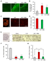
The ALX/FPR2 agonist BML-111 exerted mild attenuation of neuroinflammation 72 h after ICH induction. (A,C,E) Representative images of Iba-1 (A), MPO (C), and NeuN (E)-immunopositive cells in the peripheral region of hematoma 72 h after ICH induction. Dashed lines are the edge of the hematoma. Scale bar = 100 μm (A,C), 50 µm (E), 1 mm in low-magnification images (C,E). (B,D,F) Quantification of the Iba-1-immunopositive region in the images (640 × 640 μm2) (B), the number of MPO-immunopositive cells in the images (320 × 320 μm2) (D), and the number of NeuN-immunopositive cells in the images (230 × 190 μm2) (F). ** p < 0.01, *** p < 0.001 compared to those in the sham-operated group. Mean ± S.E.M., n = 7–9 (B), 7 (D), and 8–9 (F). Data were analyzed by one-way ANOVA with Tukey’s post hoc multiple comparisons tests. (G) Quantitative analysis of mRNA expression levels of TNF-α in the ipsilateral 4 mm thickness brain tissues containing hematoma at 72 h after ICH induction. Mean ± S.E.M., n = 4–6. ** p < 0.01 compared with the ICH group. Data were statistically analyzed by one-way ANOVA followed by Tukey’s multiple comparisons test.

References

    1. Marietta M., Pedrazzi P., Girardis M., Torelli G. Intracerebral haemorrhage: An often neglected medical emergency. Intern. Emerg. Med. 2007;2:38–45. doi: 10.1007/s11739-007-0009-y. - DOI - PMC - PubMed
    1. Keep R.F., Hua Y., Xi G. Intracerebral haemorrhage: Mechanisms of injury and therapeutic targets. Lancet Neurol. 2012;11:720–731. doi: 10.1016/S1474-4422(12)70104-7. - DOI - PMC - PubMed
    1. Zhao W., Wu C., Stone C., Ding Y., Ji X. Treatment of intracerebral hemorrhage: Current approaches and future directions. J. Neurol. Sci. 2020;416:117020. doi: 10.1016/j.jns.2020.117020. - DOI - PubMed
    1. Duan X., Wen Z., Shen H., Shen M., Chen G. Intracerebral Hemorrhage, Oxidative Stress, and Antioxidant Therapy. Oxid. Med. Cell. Longev. 2016;2016:1203285. doi: 10.1155/2016/1203285. - DOI - PMC - PubMed
    1. Bi R., Fang Z., You M., He Q., Hu B. Microglia Phenotype and Intracerebral Hemorrhage: A Balance of Yin and Yang. Front. Cell. Neurosci. 2021;15:765205. doi: 10.3389/fncel.2021.765205. - DOI - PMC - PubMed

LinkOut - more resources