. 2022 Feb 21;12(2):296.
doi: 10.3390/brainsci12020296.

Effects of Tongue Pressure on Cerebral Blood Volume Dynamics: A Functional Near-Infrared Spectroscopy Study

Affiliations

Effects of Tongue Pressure on Cerebral Blood Volume Dynamics: A Functional Near-Infrared Spectroscopy Study

Hidemasa Miyata et al. Brain Sci. .

Abstract

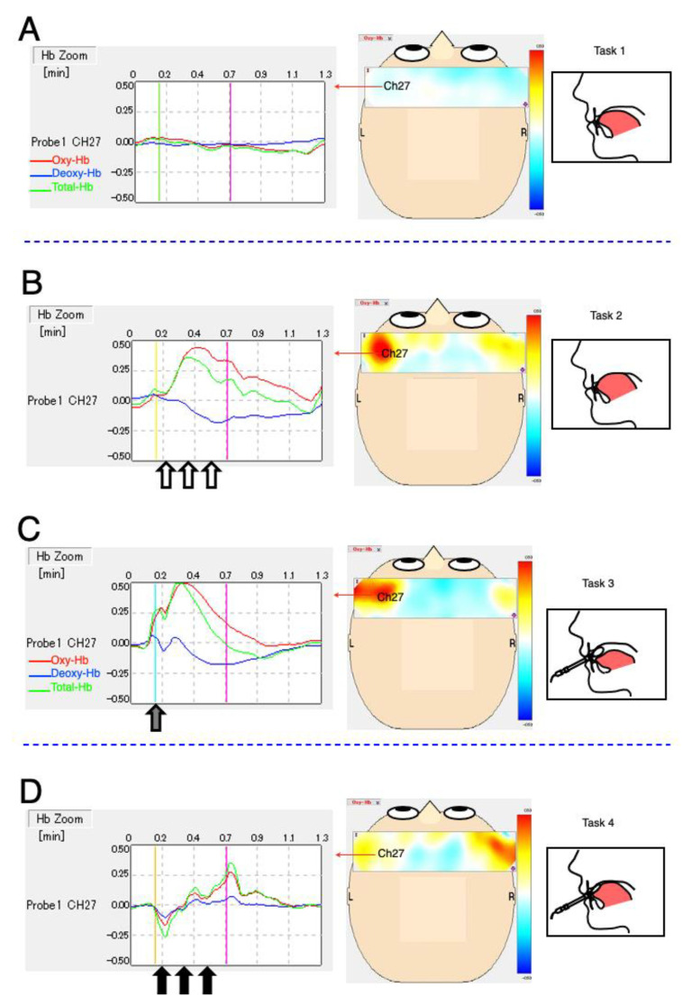
Tongue pressure measurement (TPM) is an indicator of oral function. However, the association between tongue pressure and cerebral activation remains unclear. We used near-infrared spectroscopy (NIRS) to examine the correlation between cerebral cortex activation and tongue pressure stimulation against the anterior palatal mucosa. We measured voluntary maximum tongue pressure (MTP) using a TPM device; a pressure value of approximately 60% of the MTP was used for the experimental tongue pressure (MTP60%). We examined the effect of oral functional tongue pressure stimulation against the anterior palatal mucosa on cerebral activation using NIRS in 13 adults. Tongue pressure stimulation caused significant changes in cerebral blood flow in some areas compared with controls (p < 0.05). We performed a correlation analysis (p < 0.05) between MTP60% and changes in oxygenated hemoglobin in all 47 NIRS channels. MTP60% triggered activation of the right somatosensory motor area and right dorsolateral prefrontal cortex and deactivation of the anterior prefrontal cortex (APFC). TPM balloon-probe insertion in the oral cavity activated the bilateral somatosensory motor area and deactivated the wide area of the APFC. Moreover, MTP60% via the TPM balloon probe activated the bilateral somatosensory and motor cortex areas. Tongue pressure stimulation changes cerebral blood flow, and NIRS is useful in investigating the relationship between oral stimulation and brain function.

Keywords: cerebral blood volume; cerebral cortex hemodynamics; near-infrared spectroscopy; tongue pressure measurement.


Conflict of interest statement

The authors declare no conflict of interest.

Figures

Figure 1
Figure 1
Task paradigm. The experimental design consisted of four 70 s task blocks. NIRS holder on the head during the four task measurements (top). Schematic depiction of the tongue posture during each task (second line). Images of the control stature, anterior palatal mucosa (white circle), TPM balloon-type probe, and TPM device (third line). The black line indicates sequential 30 s stimulation periods and 40 s rest periods (bottom). Task 1: resting tongue position for control. Task 2: MTP60% against anterior palatal mucosa; 5 s, three times (open arrow). Task 3: insertion of the TPM probe (gray arrow). Task 4: MTP60% via the TPM probe; 5 s, three times (solid arrow). NIRS, near-infrared spectroscopy; TPM, tongue pressure measurement, MTP, maximum tongue pressure; MTP60%, approximately 60% of the MTP for the experimental tongue pressure.
Figure 2
Figure 2
Channel allocation on the cerebral cortex. The encircled number represents near-infrared spectroscopy (NIRS: ETG-7100) 47 channels around the right temporal, frontal, and left temporal regions. PSC, primary sensory cortex; PMC, primary motor cortex; SMG, supramarginal gyrus; STG, superior temporal gyrus; MTG, middle temporal gyrus; DLPFC, dorsolateral prefrontal cortex; APFC, anterior prefrontal cortex; OFC, orbitofrontal cortex. Brain cortex schema was designed based on the report by Tsuzuki, et al. [23].
Figure 3
Figure 3
Typical waveform and topography. The left figures show near-infrared spectroscopy signal-integration waveforms at channel 27 around the left primary motor cortex; oxygenated hemoglobin (Oxy-Hb, red); deoxygenated hemoglobin (Deoxy-Hb, blue); and total hemoglobin (Total-Hb, green). Horizontal axis: time (min). The start of the tasks is indicated by four colors (light green, light yellow, turquoise-blue, and ocher, respectively), while the end of the tasks is indicated by pink. Vertical axis: Oxy-Hb values using arbitrary units (mM–mm). The center topography depict Oxy-Hb activation at the end of the tasks (pink) from overhead view and the range bar (±0.50 mm, mol–mm). (A) Resting tongue position for control. (B) MTP60% against the anterior palatal mucosa, 5 s, three times (open arrow). (C) Insertion of the TPM probe (gray arrow). (D) MTP60% via the TPM probe, 5 s, three times (solid arrow). MTP, maximum tongue pressure; MTP60%, approximately 60% of the MTP for the experimental tongue pressure; TPM, tongue pressure measurement.
Figure 4
Figure 4
Activation analysis. Significant maps were represented by Dunnett’s post hoc test of oxygenated hemoglobin (Oxy-Hb) comparing Tasks 2, 3, and 4 with Task 1 (control task) (n = 13; Dunnett’s test after analysis of variance corrected false discovery rate, p < 0.05). Red-colored circles indicate significant increases compared with Task 1; blue-colored circles indicate significant decreases compared with Task 1. (A) Task 2 vs. Task 1; (B) Task 3 vs. Task 1; (C) Task 4 vs. Task 1.
Figure 5
Figure 5
Correlation analysis. Functional connectivity between MTP60% values and changes in oxygenated hemoglobin (ΔOxy-Hb). White-encircled numbers represent significant, positively correlated channels (n = 13; p < 0.05). Scatter plots of MTP60% and ΔOxy-Hg deviation (bottom). MTP60%, approximately 60% of the MTP for the experimental tongue pressure.

Similar articles

Cited by

References

    1. Kim H.D., Choi J.B., Yoo S.J., Chang M.Y., Lee S.W., Park J.S. Tongue-to-palate resistance training improves tongue strength and oropharyngeal swallowing function in subacute stroke survivors with dysphagia. J. Oral Rehabil. 2017;44:59–64. doi: 10.1111/joor.12461. - DOI - PubMed
    1. Tsuga K., Yoshikawa M., Oue H., Okazaki Y., Tsuchioka H., Maruyama M., Yoshida M., Akagawa Y. Maximal voluntary tongue pressure is decreased in Japanese frail elderly persons. Gerodontology. 2012;29:e1078–e1085. doi: 10.1111/j.1741-2358.2011.00615.x. - DOI - PubMed
    1. Nakamori M., Hosomi N., Ishikawa K., Imamura E., Shishido T., Ohshita T., Yoshikawa M., Tsuga K., Wakabayashi S., Maruyama H., et al. Prediction of pneumonia in acute stroke patients using tongue pressure measurements. PLoS ONE. 2016;11:e0165837. doi: 10.1371/journal.pone.0165837. - DOI - PMC - PubMed
    1. Momose T., Nishikawa J., Watanabe T., Sasaki Y., Senda M., Kubota K., Sato Y., Funakoshi M., Minakuchi S. Effect of mastication on regional cerebral blood flow in humans examined by positron-emission tomography with 15O-labelled water and magnetic resonance imaging. Arch. Oral Biol. 1997;42:57–61. doi: 10.1016/S0003-9969(96)00081-7. - DOI - PubMed
    1. Sakuma S., Inamoto K., Higuchi N., Ariji Y., Nakayama M., Izumi M. Experimental pain in the gingiva and its impact on prefrontal cortical hemodynamics: A functional near-infrared spectroscopy study. Neurosci. Lett. 2014;575:74–79. doi: 10.1016/j.neulet.2014.05.040. - DOI - PubMed

LinkOut - more resources