

Clinicopathological Significance of Exosomal Proteins CD9 and CD63 and DNA Mismatch Repair Proteins in Prostate Adenocarcinoma and Benign Hyperplasia
- PMID: 35204378
- PMCID: PMC8871402
- DOI: 10.3390/diagnostics12020287
Clinicopathological Significance of Exosomal Proteins CD9 and CD63 and DNA Mismatch Repair Proteins in Prostate Adenocarcinoma and Benign Hyperplasia
Abstract
Introduction: Recently, it has been shown that exosomal biomarkers and DNA mismatch repair proteins (MMR) could play an important role in cancer risk stratification and prognosis assessment. The gold standard for prostate carcinoma (PCa) diagnosis is biopsy and histopathological examination. Thus, the complex evaluation of exosomal and MMR proteins could be beneficial for prostate cancer risk stratification and diagnostics. The aim of the current study was to evaluate and compare the expression of exosomal proteins CD9 and CD63 and MMR proteins in the tissue of patients with prostate benign hyperplasia (BPH) and PCa.
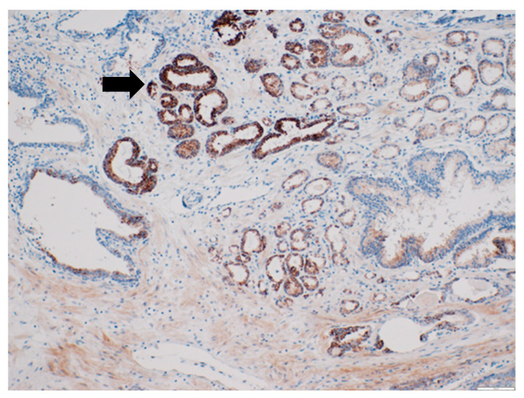
Methods: The study was retrospective. Altogether, 92 patients with PCa and 20 patients with BPH (control group) were enrolled in the study. Exosomal and MMR protein expression was analyzed by immunohistochemistry (IHC). The follow-up for each PCa patient in our study lasted till disease progression and/or a maximum of 5 years.
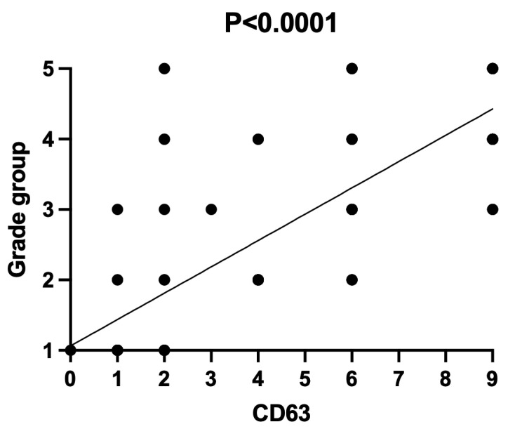
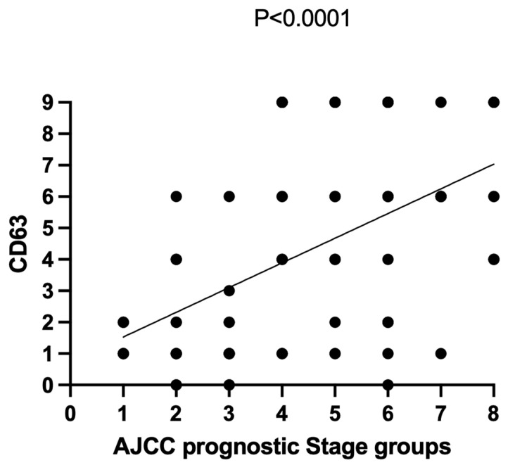
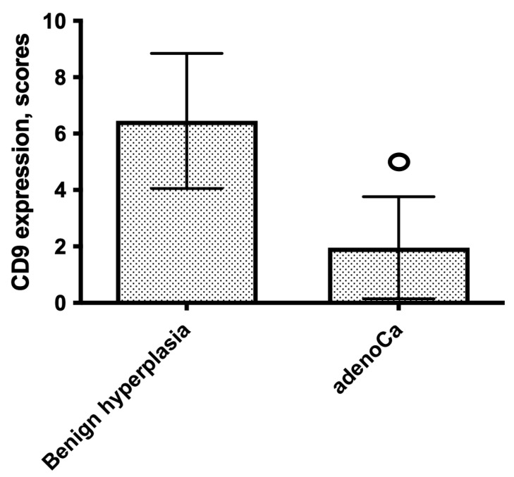
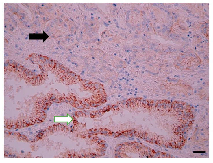
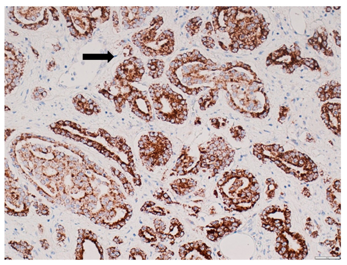
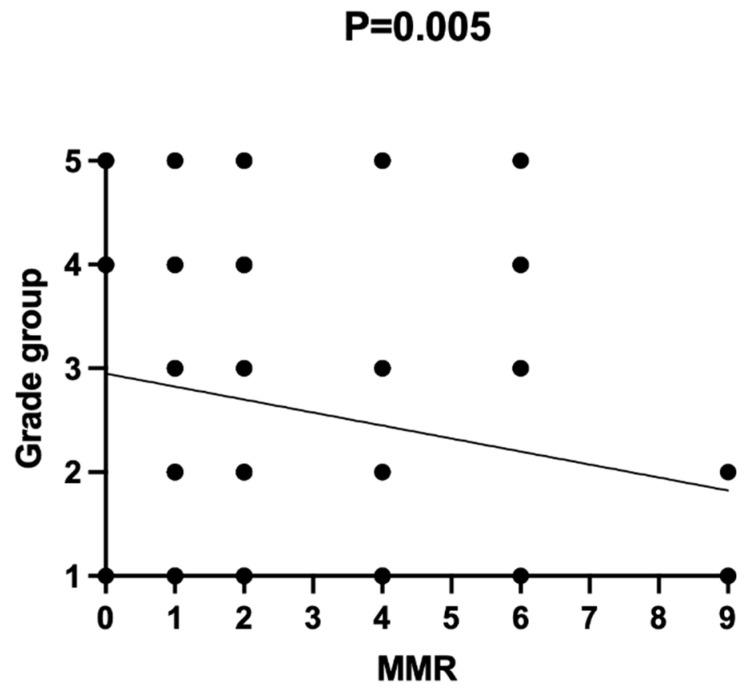
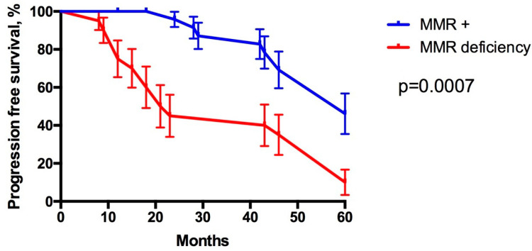
Results: Low-grade PCa was observed in 56 patients and high-grade PCa in 36 patients. CD63 expression was significantly higher in patients with high-grade PCa compared to those with low-grade PCa. CD9 expression was significantly downregulated in PCa patients compared to the control group. MMR protein expression deficiency was observed in 10 PCa patients. MMR proteins were maintained in all cases of BPH. The study found a negative correlation between MMR protein loss and PCa ISUP grade groups. Progression-free survival (PFS) in patients with MMR deficiency was significantly shorter than in patients with maintained MMR expression.
Conclusions: CD9 protein expression was downregulated in PCa, compared to BPH, while CD63 protein expression was upregulated in high-grade PCa but downregulated in low-grade PCa. CD63 protein upregulation, CD9 downregulation, and loss of MMR protein characterized the shorter PFS of high-grade PCa patients. CD9, CD63, and MMR could be the routine immunohistochemical biomarkers for the diagnosis and risk stratification of PCa.
Keywords: DNA mismatch repair proteins; benign prostate hyperplasia; exosomal biomarkers; prostate acinar adenocarcinoma.
Conflict of interest statement
The authors have no conflict of interest to declare.
Figures
References
-
- Mottet N., Cornford P., van den Bergh R.C.N., De Santis M., Fanti S., Gillessen S., Grummet J., Henry A.M., Lam T.B., Mason M.D., et al. Oncology Guidelines, Prostate Cancer. 2020. [(accessed on 9 September 2020)]. Available online: https://uroweb.org/guideline/prostate-cancer/
-
- Schmelz H.U., Sparwasser C., Weidner W. Facharztwissen Urologie, Differenzierte Diagnostik und Therapie. Springer; Heidelberg, Germnay: 2006. pp. 224–256. Kapitel 3.
-
- Yao V., Berkman C.E., Choi J.K., O’Keefe D.S., Bacich D.J. Expression of prostate-specific membrane antigen (PSMA), increases cell folate uptake and proliferation and suggests a novel role for PSMA in the uptake of the non-polyglutamated folate, folic acid. Prostate. 2010;70:305–316. doi: 10.1002/pros.21065. - DOI - PubMed
-
- Jocham D., Miller K. Praxis der Urologie. Georg Thieme Verlag; Stuttgart, Germany: 2007. pp. 128–192. Band 2, Kapitel 11.
-
- Leenders G.J.H., Kwast T.H., Grignon D.J., Evans A.J., Kristiansen G., Kweldam C.F., Litjens G., McKenney J.K., Melamed J., Mottet N., et al. The 2019 International Society of Urological Pathology (ISUP) Consensus Conference on Grading of Prostatic Carcinoma. Am. J. Surg. Pathol. 2020;44:e87–e99. doi: 10.1097/PAS.0000000000001497. - DOI - PMC - PubMed
LinkOut - more resources
Full Text Sources
Miscellaneous
