

A Challenging Diagnosis: Placental Mesenchymal Dysplasia-Literature Review and Case Report
- PMID: 35204384
- PMCID: PMC8871501
- DOI: 10.3390/diagnostics12020293
A Challenging Diagnosis: Placental Mesenchymal Dysplasia-Literature Review and Case Report
Abstract
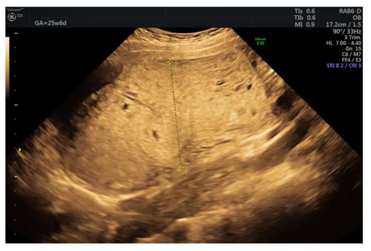
We describe a 22-year-old woman (2-gravid) case who was referred to our clinic at 18 weeks of gestation for a placenta with vesicular lesions discovered on prenatal examination routine. An ultrasound exam at 31 weeks of gestation showed numerous vesicular lesions, which gradually augmented as the pregnancy advanced. A live normal-appearing fetus was confirmed by intrauterine growth restriction (IUGR). The maternal serum β-human chorionic gonadotropin level remained in normal ranges. At some point, a multidisciplinary medical consensus considered the termination of the pregnancy, but the patient refused to comply. At 33 weeks of gestation, preterm premature rupture of membranes (pPROM) occurred, and she spontaneously delivered a 1600 g healthy female baby with a good long-term outcome. Placental mesenchymal dysplasia (PMD) was retrospectively diagnosed after confronting the data from ultrasound, chorionic villus sampling (CVS), amniocentesis, pathological examination, and immunohistochemical stain. The lack of sufficient reports of PMD determines doctors to be cautious and reserved, approaching these cases more radically than necessary. We reviewed this disease and searched for all cases of PMD associated with healthy, live newborns.
Keywords: alpha-fetoprotein; molar pregnancy; placental mesenchymal dysplasia; β-human chorionic gonadotropin.
Conflict of interest statement
The authors declare no conflict of interest.
Figures


Similar articles
-
Placental mesenchymal dysplasia: A rare case report and literature review.Medicine (Baltimore). 2025 Jun 6;104(23):e42663. doi: 10.1097/MD.0000000000042663. Medicine (Baltimore). 2025. PMID: 40489861 Free PMC article. Review.
-
Placental mesenchymal dysplasia: a case of a normal-appearing fetus with intrauterine growth restriction.Int J Clin Exp Pathol. 2014 Jul 15;7(8):5302-7. eCollection 2014. Int J Clin Exp Pathol. 2014. PMID: 25197414 Free PMC article.
-
Placental mesenchymal dysplasia, a case of intrauterine sudden death in a normal-sized fetus.J Prenat Med. 2013 Jan;7(1):9-11. J Prenat Med. 2013. PMID: 23741541 Free PMC article.
-
Placental mesenchymal dysplasia, a case of intrauterine sudden death of fetus with rupture of cirsoid periumbilical chorionic vessels.Diagn Pathol. 2011 Apr 24;6:38. doi: 10.1186/1746-1596-6-38. Diagn Pathol. 2011. PMID: 21513565 Free PMC article.
-
Diagnosis and Management of Placental Mesenchymal Disease. A Review of the Literature.Obstet Gynecol Surv. 2019 Oct;74(10):611-622. doi: 10.1097/OGX.0000000000000716. Obstet Gynecol Surv. 2019. PMID: 31670834 Review.
Cited by
-
Placental mesenchymal dysplasia: A rare case report and literature review.Medicine (Baltimore). 2025 Jun 6;104(23):e42663. doi: 10.1097/MD.0000000000042663. Medicine (Baltimore). 2025. PMID: 40489861 Free PMC article. Review.
-
Placental Mesenchymal Dysplasia With Normal Placental Growth Factor Levels Complicated by Fetal Giant Liver Cyst and Anemia.Cureus. 2025 Mar 28;17(3):e81377. doi: 10.7759/cureus.81377. eCollection 2025 Mar. Cureus. 2025. PMID: 40296964 Free PMC article.
-
Evaluation and analysis of influencing factors of placental mesenchymal dysplasia diagnosed using prenatal ultrasonography and pregnancy outcomes: case series.Quant Imaging Med Surg. 2024 Sep 1;14(9):6934-6944. doi: 10.21037/qims-23-1548. Epub 2024 Aug 28. Quant Imaging Med Surg. 2024. PMID: 39281144 Free PMC article.
-
Placental mesenchymal disease masquerading as molar pregnancy with a favourable maternal and fetal outcome.BMJ Case Rep. 2024 May 9;17(5):e258296. doi: 10.1136/bcr-2023-258296. BMJ Case Rep. 2024. PMID: 38724211
References
-
- Guenot C., Kingdom J., De Rham M., Osterheld M., Keating S., Vial Y., Van Mieghem T., Jastrow N., Raio L., Spinelli M., et al. Placental mesenchymal dysplasia: An underdiagnosed placental pathology with various clinical outcomes. Eur. J. Obstet. Gynecol. Reprod. Biol. 2019;234:155–164. doi: 10.1016/j.ejogrb.2019.01.014. - DOI - PubMed
Publication types
LinkOut - more resources
Full Text Sources
