Review
. 2022 Jan 25;12(2):306.
doi: 10.3390/diagnostics12020306.

The Diagnostic Deceiver: Radiological Pictorial Review of Tuberculosis

Affiliations
Review

The Diagnostic Deceiver: Radiological Pictorial Review of Tuberculosis

Sultan Abdulwadoud Alshoabi et al. Diagnostics (Basel). .

Abstract

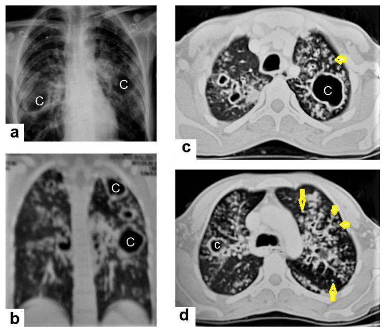
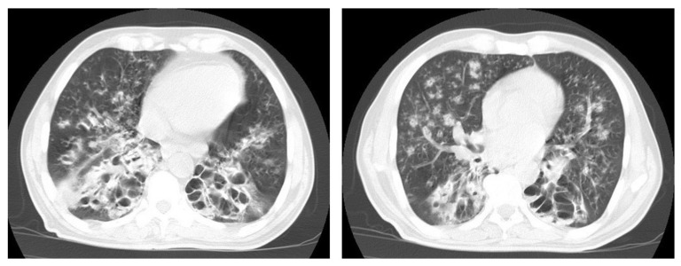
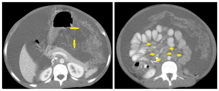
Tuberculosis (TB) is a bacterial infection with Mycobacterium tuberculosis; it is a public health problem worldwide and one of the leading causes of mortality. Since December 2019, the COVID-19 pandemic has created unprecedented health challenges and disrupted the TB health services, especially in high-burden countries with ever-increasing prevalence. Extrapulmonary and even pulmonary TB are an important cause of nonspecific clinical and radiological manifestations and can masquerade as any benign or malignant medical case, thus causing disastrous conditions and diagnostic dilemmas. Clinical manifestations and routine laboratory tests have limitations in directing physicians to diagnose TB. Medical-imaging examinations play an essential role in detecting tissue abnormalities and early suspecting diagnosis of TB in different organs. Radiologists and physicians should be familiar with and aware of the radiological manifestations of TB to contribute to the early suspicion and diagnosis of TB. The purpose of this article is to illustrate the common radiologic patterns of pulmonary and extrapulmonary TB. This article will be beneficial for radiologists, medical students, chest physicians, and infectious-disease doctors who are interested in the diagnosis of TB.

Keywords: cavitary lesions; miliary tuberculosis; tree-in-bud pattern; tuberculomas; tuberculous lymphadenitis; tuberculous pericarditis; tuberculous spondylitis.


Conflict of interest statement

The authors declare no conflict of interest.

Figures

Figure 1
Figure 1
Primary TB in an 18-year-old man. Axial mediastinal-window CT image shows multiple enlarged mediastinal lymph nodes (short arrows), and right hilar lymph nodes are characterized by central low density and peripheral enhancement after contrast administration forming the rim sign (long arrows).
Figure 2
Figure 2
Active TB in a 27-year-old man with extensive endobronchial spread. Selected axial images of chest CT show extensive endobronchial spread characterized by patchy consolidations (long arrows) in the right middle and left lower lobes with tree-in-bud nodules (short arrow) more involved the left lung. Mild right pleural effusion and mild pericardial effusion appear in both images.
Figure 3
Figure 3
Post primary TB in a 45-year-old male with cough and hemoptysis. Axial images of chest CT show (a) cavitary lesions in the right upper lobe and upper segment of the left lower lobe surrounded with consolidation, (b) cavitary lesion in the upper segment of the left lower lobe with thick irregular wall surrounded by patchy ground glass opacities, (c) centrilobular nodules and tree-in-bud nodules, and (d) consolidation in the left lower lobe with air-bronchograms.
Figure 4
Figure 4
Reactivated TB in a 34-year-old women. (a) Chest radiograph shows extensive reticular shadowing and multiple cavitary lesions in both lungs. (b) Coronal reconstruction CT shows multiple cavitary lesions (c) in both lungs, the large two in the posterior segment of the upper lobe, and in the apical segment of the lower lobe of the left lung. (c,d) Axial CT images of the lung show multiple cavitary lesion, the largest in the posterior segment of the left upper lobe, with multiple centrilobular (long arrows) and tree-in-bud (short arrows) appearance.
Figure 5
Figure 5
Active TB in a 27-year-old man (same case of Figure 2) with endobronchial spread. (a) Axial image of chest CT shows endobronchial spread characterized centrilobular and tree-in-bud nodules involved the upper lobe of the left lung. (b) CT axial image with mediastinal window shows large necrotic mediastinal lymph node measures 21 mm × 20 mm (arrow) in the anterior mediastinum.
Figure 6
Figure 6
Miliary TB in an 18-year-old man (same patient of Figure 1). (a) Chest radiograph shows superimposed innumerable small nodules in lungs right greater than the left predominantly affecting lung bases due to gravity-dependent high blood flow. (b) Axial chest CT shows innumerable small (1–3 mm) nodules with random distribution in both lung fields.
Figure 7
Figure 7
Miliary TB in an adult female with fatigue and loss of appetite. (a) Splenic ultrasonography shows multiple small hypoechoic granulomas with random distribution in the spleen. (b) Axial abdominal CT shows small non-enhancing granulomas with random distribution in the spleen clearly seen during the portal venous phase with multiple enlarged lymph nodes around the portal vein.
Figure 8
Figure 8
Active TB in a 27-year-old man (same case of Figure 2 and Figure 5). (a) Axial image of chest CT shows moderate left and mild right pleural effusion with discrete pulmonary nodules in active TB. (b) Axial image of the mediastinal window shows moderate left and mild right pleural effusion with mild pericardial effusion.
Figure 9
Figure 9
Old reactive TB with superimposed infection in a 75-year-old female with chronic cough and no history of previous medication. Selected axial images of lung CT show prominent fibrotic changes with scarring traction bronchiectasis and decreased volume in the apical and posterior segment of the upper lobes and architectural distortion of the lung parenchyma caused by cystic bronchiectasis predominantly involve bilateral lower lobes, ligula, and right middle lobe. Patchy consolidative areas and ground glass opacities are signs of active infection.
Figure 10
Figure 10
Reactivation of TB in an adult man with chronic cough. Selected axial images of lung CT show cystic bronchiectasis, due to old infection, with peribronchial thickening and air fluid level involving both lower lobes. Scattered tree in bud pattern and infected cystic bronchiectasis indicate active TB.
Figure 11
Figure 11
Post-primary-TB in a 45-year-old man. Selected axial and coronal reconstruction images of chest CT show multiple thick-walled cavitary lesions (arrows) in both lungs, with left pneumothorax as a complication of TB. Surgical emphysema in the left chest wall, due to chest-tube insertion.
Figure 12
Figure 12
Tuberculous leptomeningitis. Selected axial T1-weighted images of brain MRI post-contrast administration show diffuse leptomeningeal enhancement (short arrows) with predilection to involve the basal cisterns complicated with hydrocephalus. Multiple ring-enhancing lesions (long arrows) in different regions of the brain.
Figure 13
Figure 13
Multiple tuberculomas in an adult man. Selected MRI axial images post-contrast T1-weighted images show multiple small ring-enhancing lesions distributed in both cerebral hemispheres, with no significant surrounding cerebral edema, as is consistent with cerebral tuberculomas.
Figure 14
Figure 14
Tuberculous cerebritis in an adult man with fever and seizures. (a) Axial FLAIR-weighted images of brain MRI show high signal intensity in the right parietal and frontal lobes causing mass effect compressing the right lateral ventricle with mild shifting of the midline to the contralateral side. (b) Axial brain T1-weighted images post-contrast administration show intense serpentine (gyriform) enhancement in the right parietal and frontal lobes, with significant surrounding edema, suggesting tuberculous cerebritis.
Figure 15
Figure 15
Tuberculous abscesses and tuberculomas in a 70-year-old woman with disturbance of consciousness and long history of headache. Selected axial computed tomography (CT) images of the brain (a) axial non-enhanced CT show significant vasogenic edema in the frontal and parietal lobes of the right cerebral hemisphere, with severe mass effect manifested as loss of the cortical sulci, total effacement of the right lateral ventricle and mild midline shift to the left side. (b) Contrast-enhanced CT (CECT) shows loculated rim-enhancing lesion centered in the right frontal lobe measures about 39 mm × 16 mm × 12 mm, suggesting cerebral abscess (arrows). (c) Axial CECT shows multiple ring-enhancing lesions in the right cerebral hemispheres and right basal ganglia (short arrows) suggesting of cerebral abscesses or tuberculomas. (d) Coronal CECT shows multiple ring-enhancing lesions in the right cerebral hemispheres (short arrows), suggesting cerebral tuberculomas or abscesses.
Figure 16
Figure 16
Tuberculous abscess in the right cerebrum of a 13-year-old female. Selected images of brain MRI (a) T1-weighted image (WI), (b) T2-WIs, (c) T1-WI with contrast, and (d) FLAIR-WI. The images show a well-defined lesion in the right cerebral hemisphere with low-signal-intensity content and a high-signal-intensity capsule on T1WI, which appear as high-signal-intensity content and a low-signal-intensity capsule on T2WI (b); there is marginal enhancement on T1WI with contrast administration (c), and there are low-signal-intensity contents and a high-signal-intensity capsule on FLAIR (d). Obvious grade-2 vasogenic oedema around the lesion on T2WI and FLAIR.
Figure 17
Figure 17
A tuberculous abscess in the cerebellum of a 13-year-old female (same patient as Figure 16). Selected images of brain MRI (a) T1-weighted image (WI), (b) FLAIR-WIs, (c) T1-WI with contrast, and (d) diffusion-weighted (DW) image. The images show a well-defined lesion in the cerebellum with low signal intensity content and a high signal intensity capsule on T1WI and FLAIR (b), marginal enhancement on T1WI with contrast administration (c), and restricted diffusion on DWI (d).
Figure 18
Figure 18
Pott’s disease in a 28-year-old female. Selected images of sagittal MRI of thoracic spine show destructive lesion affects multiple contiguous thoracic vertebrae with paraspinal collection. (a) Pre-contrast and (b) post-contrast T1-weighted images show subligamentous spread of the infection to involve five vertebrae with peripheral enhancement of the lesions, suggesting an abscess (arrow).
Figure 19
Figure 19
Pott’s disease in a 6-year-old female patient. Selected images of lumbosacral MRI. Sagittal (a) T1-weighted images and (b) T2-weighted images show that destructive lesion involves the T4/T5 disc and vertebrae, with extensive pre-vertebral loculated fluid collection. Selected axial images of MRI (c) T1-weighted image with gadolinium, and (d) T2-weighted image shows bilateral paravertebral loculated fluid collections involved in bilateral psoas; paravertebral muscles appear to have low-signal-intensity contents on T1-WIs with marginal enhancement after contrast administration and high-signal-intensity contents on T2-WIs (arrows). The picture is typical of Pott’s disease with bilateral psoas and paravertebral abscesses.
Figure 20
Figure 20
Tuberculous lymphadenitis in a 27-year-old women with cachexia, loss of appetite and cough. Axial abdominal CT image shows enlarged mesenteric, periaortic, and portahepatis lymph nodes (LNs), (short arrows) due to TB, which usually involves multiple groups, such as mesenteric and upper paraortic LNs. The image also shows relatively dense ascites and remarkable omental thickening forming cake-like mass (long arrow).
Figure 21
Figure 21
Wet peritonitis in a 16-year-old man with cachexia and loss of appetite. Selected axial abdominal images show marked relatively dense ascites and gross thickened omentum with faint enhancement of peritoneal reflections (long arrows) with multiple enlarged mesenteric and upper paraortic conglomerated lymph nodes (short arrows) with slight homogeneous enhancement.
Figure 22
Figure 22
Fibrotic peritonitis in a 20-year-old man with fatigue, abdominal distension, and loss of appetite. Selected axial images of abdominal CT show remarkable omental thickening, forming cake-like masses (arrows) with faint enhancement and mild smooth thickening of peritoneal reflections associated with mild-to-moderate dense ascites and several low-attenuation mesenteric lymph nodes that are challenging to be distinguished from small bowel loops.
Figure 23
Figure 23
Renal TB in a 39-year-old man. Selected axial computed tomography images show (a) focal hyperdense non-enhancing nodules, with the largest at 26 mm, with calcified rim in the upper pole of the right kidney with focal scarring of the kidney. (b,c) Approximately, 20 mm cystic lesion in the upper calyx with focal calcification, most likely dilated calyx; and (d) 11 mm focal parenchymal calcification in the middle calyx.
Figure 24
Figure 24
Knee TB in a 45-year-old patient with proved case of knee TB infection. Selected sagittal MRI images of (a) T1-weighted image and (b) T2-weighted image show extensive oedema of the articular surfaces of the knee, appearing as low signal intensity on T1 and high signal intensity on T2 with diffuse synovial thickening and large bone erosions (arrow heads), with mild joint effusion and a prepatellar pocket of fluid collection, which appear as low signal intensity on T1 and high signal intensity on T2. In addition, multiple enlarged popliteal lymph nodes were present.

References

    1. Nachiappan A.C., Rahbar K., Shi X., Guy E.S., Mortani Barbosa E.J., Jr., Shroff G.S., Ocazionez D., Schlesinger A.E., Katz S.I., Hammer M.M. Pulmonary Tuberculosis: Role of Radiology in Diagnosis and Management. Radiographics. 2017;37:52–72. doi: 10.1148/rg.2017160032. - DOI - PubMed
    1. Sahu S., Ditiu L., Sachdeva K.S., Zumla A. Recovering from the Impact of the COVID-19 Pandemic and Accelerating to Achieving the United Nations General Assembly Tuberculosis Targets. Int. J. Infect. Dis. 2021;S1201–S9712:164–168. doi: 10.1016/j.ijid.2021.02.078. - DOI - PMC - PubMed
    1. Ringold S., Lynm C., Glass R.M. Tuberculosis. JAMA. 2005;293:2820. doi: 10.1001/jama.293.22.2820. - DOI - PubMed
    1. Lee S.H. Tuberculosis Infection and Latent Tuberculosis. Tuberc. Respir. Dis. 2016;79:201–206. doi: 10.4046/trd.2016.79.4.201. - DOI - PMC - PubMed
    1. Tan C.H., Kontoyiannis D.P., Viswanathan C., Iyer R.B. Tuberculosis: A benign impostor. AJR Am. J. Roentgenol. 2010;194:555–561. doi: 10.2214/AJR.09.3055. - DOI - PubMed

LinkOut - more resources