

Metallic Component Preserving Algorithm Based on the Cerebral Computed Tomography Angiography in Aneurysm Surgery
- PMID: 35204429
- PMCID: PMC8871085
- DOI: 10.3390/diagnostics12020338
Metallic Component Preserving Algorithm Based on the Cerebral Computed Tomography Angiography in Aneurysm Surgery
Abstract
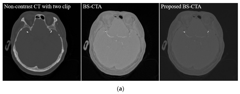
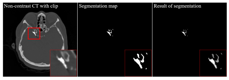
The purpose of this study was to investigate the viability of the proposed method in preventing the loss of metallic components including the clip and coil in cerebral computed tomography angiography (CTA). Forty patients undergoing surgery for aneurysms carried metallic materials. The proposed method is based on conventional bone subtraction CTA (BS-CTA) system. Briefly, the position of metal components was determined using the threshold value and a region of interest (ROI). An appropriate threshold was used to separate the background from the target materials based on the Otsu method. A three-dimensional (3D) rendering was performed from the proposed BS-CTA data carrying the extracted target information. The accuracy of clip and coil region measured using the dice similarity coefficient (DSC) and bidirectional Hausdorff distance (HD) is reported. The metallic components of the proposed BS-CTA were significantly visualized in various patient cases. Quantitative evaluation using the proposed method is based on the mean DSC of 0.93 with a standard deviation (SD) of ±0.05 (e.g., maximum value = 0.99, minimum value = 0.75, 95% confidence interval (CI) = 0.91 to 0.95, and all p < 0.05). The mean HD was 1.50 voxels with an SD of ± 0.58 (e.g., maximum value = 5.95, minimum value = 0.12, 95% CI = 1.10 to 1.90, and all p < 0.05). The proposed method demonstrates effective segmentation of the metallic component and application to the existing conventional BS-CTA system.
Keywords: aneurysm; bone subtraction; cerebral CT angiography (CTA); clip and coil; performance evaluation.
Conflict of interest statement
The authors declare no conflict of interest.
Figures


Similar articles
-
Dual-energy CT angiography in the evaluation of intracranial aneurysms: image quality, radiation dose, and comparison with 3D rotational digital subtraction angiography.AJR Am J Roentgenol. 2010 Jan;194(1):23-30. doi: 10.2214/AJR.08.2290. AJR Am J Roentgenol. 2010. PMID: 20028901
-
Value of automatic bone subtraction in cranial CT angiography: comparison of bone-subtracted vs. standard CT angiography in 100 patients.Eur Radiol. 2008 May;18(5):974-82. doi: 10.1007/s00330-008-0855-7. Epub 2008 Jan 26. Eur Radiol. 2008. PMID: 18224325
-
Evaluation of postoperative status after clipping surgery in patients with cerebral aneurysm on 3-dimensional-CT angiography with elimination of clips.J Neuroimaging. 2011 Jan;21(1):10-5. doi: 10.1111/j.1552-6569.2009.00435.x. J Neuroimaging. 2011. PMID: 19888935
-
Detection of intracranial aneurysms using three-dimensional multidetector-row CT angiography: is bone subtraction necessary?Eur J Radiol. 2011 Aug;79(2):e18-23. doi: 10.1016/j.ejrad.2010.01.004. Epub 2010 Feb 9. Eur J Radiol. 2011. PMID: 20144517
-
[Diagnostic imaging of cerebrovascular disease on multi-detector row computed tomography (MDCT)].Brain Nerve. 2011 Sep;63(9):923-32. Brain Nerve. 2011. PMID: 21878694 Review. Japanese.
Cited by
-
Comparison of bone subtraction CT angiography with standard CT angiography for evaluating circle of Willis in normal dogs.J Vet Sci. 2023 Sep;24(5):e65. doi: 10.4142/jvs.23121. J Vet Sci. 2023. PMID: 38031644 Free PMC article.
References
LinkOut - more resources
Full Text Sources
