Review
. 2022 Feb 2;12(2):384.
doi: 10.3390/diagnostics12020384.

Imaging Review of Pelvic Ring Fractures and Its Complications in High-Energy Trauma

Affiliations
Review

Imaging Review of Pelvic Ring Fractures and Its Complications in High-Energy Trauma

Edoardo Leone et al. Diagnostics (Basel). .

Abstract

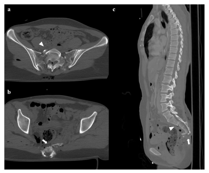
Pelvic ring fractures are common in high-energy blunt trauma, especially in traffic accidents. These types of injuries have a high rate of morbidity and mortality, due to the common instability of the fractures, and the associated intrapelvic vascular and visceral complications. Computed tomography (CT) is the gold standard technique in the evaluation of pelvic trauma because it can quickly and accurately identify pelvic ring fractures, intrapelvic active bleeding, and lesions of other body systems. To properly guide the multidisciplinary management of the polytrauma patient, a classification criterion is mandatory. In this review, we decided to focus on the Young and Burgess classification, because it combines the mechanism and the stability of the fractures, helping to accurately identify injuries and related complications.

Keywords: computed tomography; genitourinary injury; high-energy trauma; intrapelvic bleeding; pelvic ring fracture.


Conflict of interest statement

The authors declare no conflict of interest.

Figures

Figure 1
Figure 1
Lateral compression fracture, type 1. Axial computed tomography (CT) images show a fracture of the right-side superior and inferior pubic rami (arrows in (a,b)), and ipsilateral fracture of the sacrum (arrowheads in (c)); the sacral fracture involves the neuroforamina (zone II). Three-dimensional volume-rendering CT reconstruction in anteroposterior (AP) view confirms the type of fracture (d).
Figure 2
Figure 2
Lateral compression fracture, type 2. Axial CT images show a fracture of both the sacral wings (arrows in (a)), without the involvement of the neuroforamina (zone I). There is also fracture of the iliac wing on the right side (arrow in (b)), and ipsilateral fracture of the superior and inferior pubic branches (arrowheads in (c,d)).
Figure 3
Figure 3
Lateral compression fracture, type 3. Axial CT images show the fracture of the iliac and sacral wings on the left side (a), anterior widening of the right sacroiliac joint (b), and fracture of bilateral pubic branches (c,d).
Figure 4
Figure 4
Anterior–posterior compression fracture, type 1. Anteroposterior scout CT view (a) and axial CT image (b) show a 2.4 cm diastasis of the pubic symphysis. Three-dimensional volume-rendering CT reconstructions in outlet and inlet views (c,d) well depict the diastasis.
Figure 5
Figure 5
Anterior–posterior compression fracture, type 2. Axial CT images (a,b) show a pubic symphysis diastasis of 3.2 cm, and an anterior sacroiliac joint diastasis on the left side. Three-dimensional volume-rendering CT reconstructions in outlet and inlet views (c,d) confirm the findings.
Figure 6
Figure 6
Anterior–posterior compression fracture, type 3. Axial CT images (a,b) and three-dimensional volume-rendering CT reconstructions in AP and inlet views (c,d) show a pubic symphysis diastasis of 4.2 cm, anterior and posterior left side sacroiliac joint diastasis, and anterior widening of the right sacroiliac joint.
Figure 7
Figure 7
Vertical shear fracture. Axial CT images show avulsion of the left transverse process of the fifth lumbar vertebra (black arrow in (a)), fracture of the left side of the sacrum with involvement of the foraminal zone (black arrowhead in (b)), and fracture of the right superior and inferior pubic branches (white arrows in (c,d)). There is also a little fracture of the right wing of the sacrum, and a posterior widening of the right sacroiliac joint (white arrowheads in (b)). Three-dimensional volume-rendering CT reconstruction clearly depicts the instability of the pelvic ring, with the fracture of the left transverse process of the fifth lumbar vertebra and the cranial shift of the left hemipelvis (e).
Figure 8
Figure 8
Fractures of the sacrum with involvement of the central canal. Fracture through S2, with anterior angulation of the superior fragment and without dislocation (arrowheads in (a,c)). Fracture of S4 with complete anterolisthesis of the fragments (arrows in (b,c)). There are also some bubbles of air in the adjacent soft tissues.
Figure 9
Figure 9
Plain radiography. The outlet view (a) and the inlet view (b) show a 5.2 cm diastasis of the pubic symphysis and anterior widening of the left-side sacroiliac joint. These findings have to be quickly reported to allow prompt treatment.
Figure 10
Figure 10
Pelvic ring trauma, characterized by fracture of right superior and inferior pubic rami. Axial CT image in the arterial phase shows active extravasation of contrast medium near the right superior pubic branch (arrow in (a)), which increases in the venous phase of the examination (arrow in (b)). Similar active extravasation is documented near the internal obturator muscle (arrowheads in (d,e)). Selective angiography confirmed the two blood extravasation spots, with origin from branches of the right obturator artery (arrows and arrowhead in (c,f)); both the bleeding spots were optimally embolized.
Figure 11
Figure 11
Axial CT images show a pubic symphysis diastasis of 2.9 cm, and an anterior widening of the right sacroiliac joint (a,b). Active extravasation of contrast medium in the arterial phase of the CT exam is seen near the right side of the fifth sacral vertebra (arrow in (c)). The extravasation increases in the next phase of the examination (arrow in (d)). This is acute arterial bleeding from a branch of the right inferior gluteal artery.
Figure 12
Figure 12
Patient of the Figure 5, with an anterior–posterior compression fracture. There is a large hematoma inside the bladder (a); contrast medium administration reveals active bleeding in the venous phase of the examination, on the left wall of the bladder (b). Axial and coronal CT cystography images show a leak of contrast medium in the perivesical space, suggestive of extraperitoneal bladder rupture (c,d).
Figure 13
Figure 13
Patient of the Figure 6, with an extraperitoneal bladder rupture. The retrograde introduction of air shows a leak at the level of the anterior wall (a,b).
Figure 14
Figure 14
Three-dimensional volume-rendering CT reconstructions in AP and inlet views show an anterior–posterior compression fracture (a,b). Coronal CT image shows hematoma in the right inguinoscrotal region (c). Ultrasound shows an inhomogeneous hematoma on the extra-albuginea side (arrow in (d,e)) and a hypoechoic area in the upper part of the testis (star in (e)). Contrast-enhanced ultrasonography (CEUS) image clearly shows that the hypoechoic area is avascularized, as a hematoma (star in (f)). Ultrasound and CEUS also show the irregularity of the testicular contour (arrowhead in (e,f)). These findings are suggestive of testicular rupture.

References

    1. Grotz M.R.W., Allami M.K., Harwood P., Pape H.C., Krettek C., Giannoudis P.V. Open pelvic fractures: Epidemiology, current concepts of management and outcome. Injury. 2005;36:1–13. doi: 10.1016/j.injury.2004.05.029. - DOI - PubMed
    1. Magnone S., Coccolini F., Manfredi R., Piazzalunga D., Agazzi R., Arici C., Barozzi M., Bellanova G., Belluati A., Berlot G., et al. Management of hemodynamically unstable pelvic trauma: Results of the first Italian consensus conference (cooperative guidelines of the Italian Society of Surgery, the Italian Association of Hospital Surgeons, the Multi-specialist Italian Society of Young Surgeons, the Italian Society of Emergency Surgery and Trauma, the Italian Society of Anesthesia, Analgesia, Resuscitation and Intensive Care, the Italian Society of Orthopaedics and Traumatology, the Italian Society of Emergency Medicine, the Italian Society of Medical Radiology -Section of Vascular and Interventional Radiology- and the World Society of Emergency Surgery) World. J. Emerg. Surg. 2014;9:9–18. doi: 10.1186/1749-7922-9-18. - DOI - PMC - PubMed
    1. Coccolini F., Stahel P.F., Montori G., Biffl W., Horer T.M., Catena F., Kluger Y., Moore E.E., Peitzman A.B., Ivatury R., et al. Pelvic trauma: WSES classification and guidelines. World J. Emerg Surg. 2017;12:5. doi: 10.1186/s13017-017-0117-6. - DOI - PMC - PubMed
    1. McCormak R., Strauss E.J., Alwatter B.J., Tejwani N.C. Diagnosis and management of pelvic fractures. Bull. NYU Hosp. Jt. Dis. 2010;68:281–291. - PubMed
    1. Yoshihara H., Yoneoka D. Demographic epidemiology of unstable pelvic fracture in the United States from 2000 to 2009: Trends and in-hospital mortality. J. Trauma Acute Care Surg. 2014;76:380–385. doi: 10.1097/TA.0b013e3182ab0cde. - DOI - PubMed

LinkOut - more resources