

A CT-Based Radiomic Signature for the Differentiation of Pulmonary Hamartomas from Carcinoid Tumors
- PMID: 35204507
- PMCID: PMC8871366
- DOI: 10.3390/diagnostics12020416
A CT-Based Radiomic Signature for the Differentiation of Pulmonary Hamartomas from Carcinoid Tumors
Abstract
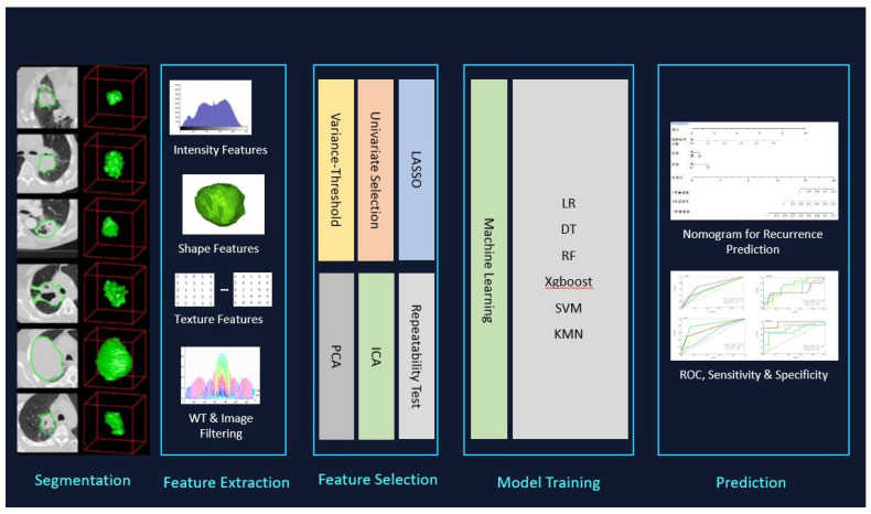
Radiomics is a new image processing technology developed in recent years. In this study, CT radiomic features are evaluated to differentiate pulmonary hamartomas (PHs) from pulmonary carcinoid tumors (PCTs). A total of 138 patients (78 PCTs and 60 PHs) were evaluated. The Radcloud platform (Huiying Medical Technology Co., Ltd., Beijing, China) was used for managing the data, clinical data, and subsequent radiomics analysis. Two hand-crafted radiomics models are prepared in this study: the first model includes the data regarding all of the patients to differentiate between the groups; the second model includes 78 PCTs and 38 PHs without signs of fat tissue. The separation of the training and validation datasets was performed randomly using an (8:2) ratio and 620 random seeds. The results revealed that the MLP method (RF) was best for PH (AUC = 0.999) and PCT (AUC = 0.999) for the first model (AUC = 0.836), and PC (AUC = 0.836) in the test set for the second model. Radiomics tumor features derived from CT images are useful to differentiate the carcinoid tumors from hamartomas with high accuracy. Radiomics features may be used to differentiate PHs from PCTs with high levels of accuracy, even without the presence of fat on the CT. Advances in knowledge: CT-based radiomic holds great promise for a more accurate preoperative diagnosis of solitary pulmonary nodules (SPNs).
Keywords: carcinoid; machine learning; pulmonary hamartomas; radiomics.
Conflict of interest statement
The authors declare no conflict of interest.
Figures


References
-
- De Cicco C., Bellomi M., Bartolomei M., Carbone G., Pelosi G., Veronesi G., De Pas T., Spaggiari L., Paganelli G. Imaging of lung hamartomas by multidetector computed tomography and positron emission tomography. Ann. Thorac. Surg. 2008;86:1769–1772. doi: 10.1016/j.athoracsur.2008.08.033. - DOI - PubMed
LinkOut - more resources
Full Text Sources
