

Inferior Vena Cava Edge Tracking Echocardiography: A Promising Tool with Applications in Multiple Clinical Settings
- PMID: 35204518
- PMCID: PMC8871248
- DOI: 10.3390/diagnostics12020427
Inferior Vena Cava Edge Tracking Echocardiography: A Promising Tool with Applications in Multiple Clinical Settings
Abstract
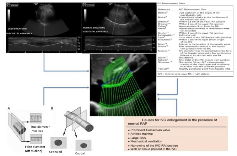
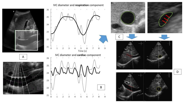
Ultrasound (US)-based measurements of the inferior vena cava (IVC) diameter are widely used to estimate right atrial pressure (RAP) in a variety of clinical settings. However, the correlation with invasively measured RAP along with the reproducibility of US-based IVC measurements is modest at best. In the present manuscript, we discuss the limitations of the current technique to estimate RAP through IVC US assessment and present a new promising tool developed by our research group, the automated IVC edge-to-edge tracking system, which has the potential to improve RAP assessment by transforming the current categorical classification (low, normal, high RAP) in a continuous and precise RAP estimation technique. Finally, we critically evaluate all the clinical settings in which this new tool could improve current practice.
Keywords: caval index; edge tracking; heart failure; inferior vena cava; pulmonary hypertension; right atrial pressure.
Conflict of interest statement
The authors declare no conflict of interest.
Figures


Similar articles
-
Non-Invasive Estimation of Right Atrial Pressure Using a Semi-Automated Echocardiographic Tool for Inferior Vena Cava Edge-Tracking.J Clin Med. 2022 Jun 7;11(12):3257. doi: 10.3390/jcm11123257. J Clin Med. 2022. PMID: 35743330 Free PMC article.
-
Processing Ultrasound Scans of the Inferior Vena Cava: Techniques and Applications.Bioengineering (Basel). 2023 Sep 12;10(9):1076. doi: 10.3390/bioengineering10091076. Bioengineering (Basel). 2023. PMID: 37760178 Free PMC article. Review.
-
Accuracy of right atrial pressure estimation using a multi-parameter approach derived from inferior vena cava semi-automated edge-tracking echocardiography: a pilot study in patients with cardiovascular disorders.Int J Cardiovasc Imaging. 2020 Jul;36(7):1213-1225. doi: 10.1007/s10554-020-01814-8. Epub 2020 Mar 19. Int J Cardiovasc Imaging. 2020. PMID: 32193772
-
Correlation between right atrial pressure measured via right heart catheterization and venous excess ultrasound, inferior vena cava diameter, and ultrasound-measured jugular venous pressure: a prospective observational study.Ultrasound J. 2024 Nov 29;16(1):50. doi: 10.1186/s13089-024-00397-y. Ultrasound J. 2024. PMID: 39612127 Free PMC article.
-
The efficacy of sonographic measurement of inferior vena cava diameter as an estimate of central venous pressure.Cardiovasc Ultrasound. 2016 Aug 20;14(1):33. doi: 10.1186/s12947-016-0076-1. Cardiovasc Ultrasound. 2016. PMID: 27542597 Free PMC article. Review.
Cited by
-
Non-Invasive Estimation of Right Atrial Pressure Using a Semi-Automated Echocardiographic Tool for Inferior Vena Cava Edge-Tracking.J Clin Med. 2022 Jun 7;11(12):3257. doi: 10.3390/jcm11123257. J Clin Med. 2022. PMID: 35743330 Free PMC article.
-
Refining accuracy of RV-PA coupling in patients undergoing transcatheter tricuspid valve treatment.Clin Res Cardiol. 2024 Jan;113(1):177-186. doi: 10.1007/s00392-023-02339-5. Epub 2023 Nov 27. Clin Res Cardiol. 2024. PMID: 38010521 Free PMC article.
-
Evaluation of an artificial intelligence-based system for echocardiographic estimation of right atrial pressure.Int J Cardiovasc Imaging. 2023 Dec;39(12):2437-2450. doi: 10.1007/s10554-023-02941-8. Epub 2023 Sep 8. Int J Cardiovasc Imaging. 2023. PMID: 37682418 Free PMC article.
-
Processing Ultrasound Scans of the Inferior Vena Cava: Techniques and Applications.Bioengineering (Basel). 2023 Sep 12;10(9):1076. doi: 10.3390/bioengineering10091076. Bioengineering (Basel). 2023. PMID: 37760178 Free PMC article. Review.
-
Prolapsing Intrigue: A Case of Superior Vena Cava Mass Visualized by Echocardiography From the Subcostal Window Unveiling an Anterior Mediastinal Type B2 Thymoma.Cureus. 2024 Jan 16;16(1):e52352. doi: 10.7759/cureus.52352. eCollection 2024 Jan. Cureus. 2024. PMID: 38230386 Free PMC article.
References
-
- Rudski L.G., Lai W.W., Afilalo J., Hua L., Handschumacher M.D., Chandrasekaran K., Solomon S.D., Louie E.K., Schiller N.B. Guidelines for the Echocardiographic Assessment of the Right Heart in Adults: A Report from the American Society of Echocardiography. Endorsed by the European Association of Echocardiography, a registered branch of the European Society of Cardiology. J. Am. Soc. Echocardiogr. 2010;23:685–713. doi: 10.1016/j.echo.2010.05.010. - DOI - PubMed
Publication types
LinkOut - more resources
Full Text Sources
