. 2022 Feb 14;12(2):495.
doi: 10.3390/diagnostics12020495.

Evaluation of a Generative Adversarial Network to Improve Image Quality and Reduce Radiation-Dose during Digital Breast Tomosynthesis

Affiliations

Evaluation of a Generative Adversarial Network to Improve Image Quality and Reduce Radiation-Dose during Digital Breast Tomosynthesis

Tsutomu Gomi et al. Diagnostics (Basel). .

Abstract

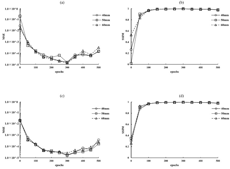
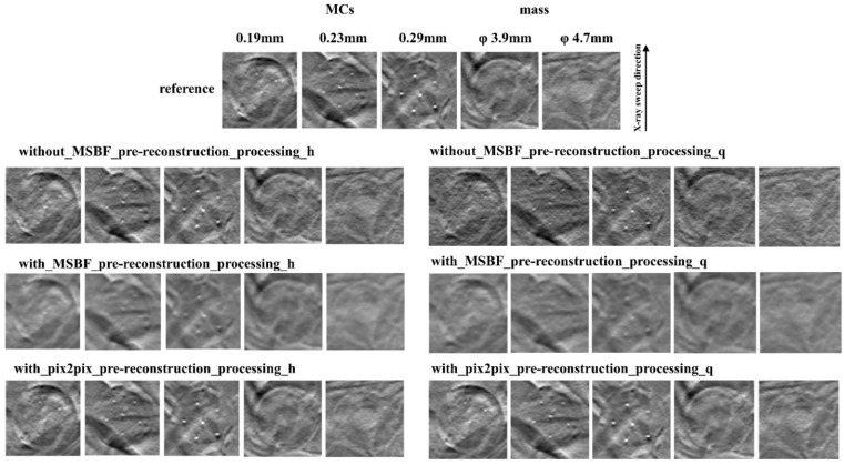
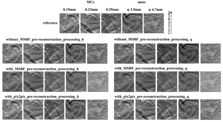
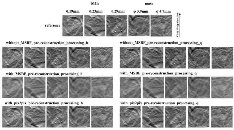
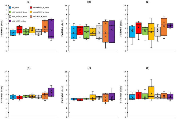
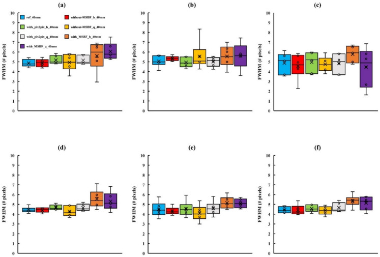
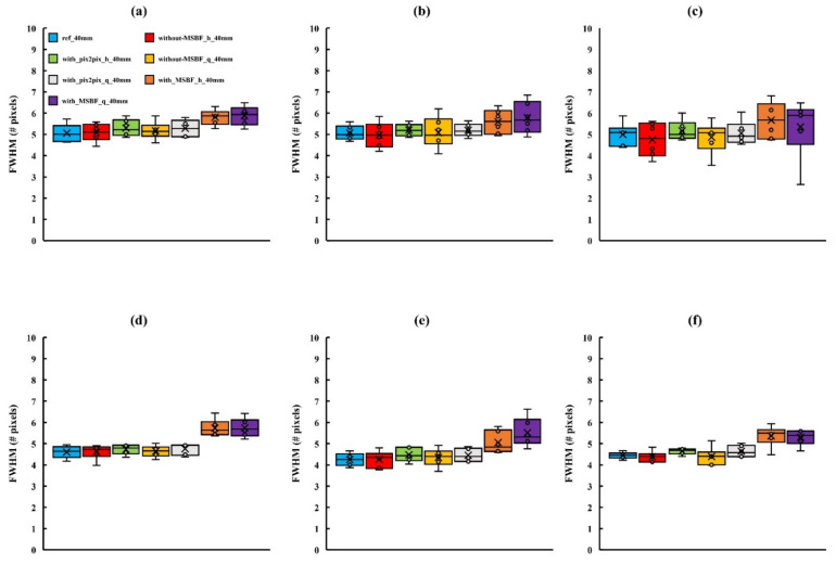
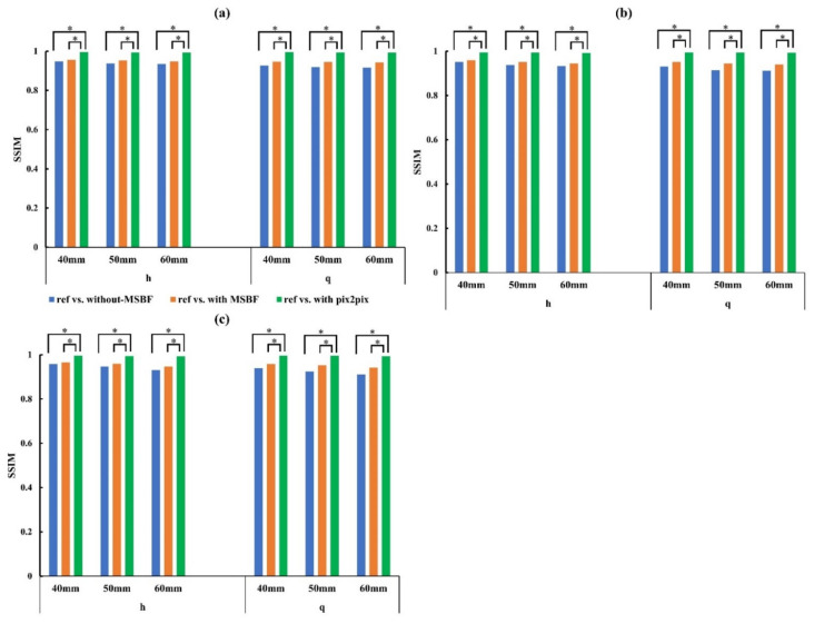
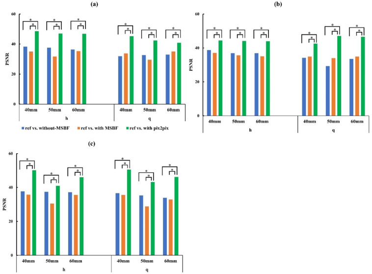
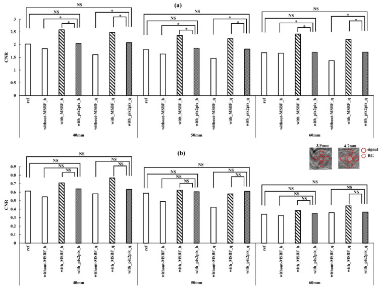
In this study, we evaluated the improvement of image quality in digital breast tomosynthesis under low-radiation dose conditions of pre-reconstruction processing using conditional generative adversarial networks [cGAN (pix2pix)]. Pix2pix pre-reconstruction processing with filtered back projection (FBP) was compared with and without multiscale bilateral filtering (MSBF) during pre-reconstruction processing. Noise reduction and preserve contrast rates were compared using full width at half-maximum (FWHM), contrast-to-noise ratio (CNR), peak signal-to-noise ratio (PSNR), and structural similarity (SSIM) in the in-focus plane using a BR3D phantom at various radiation doses [reference-dose (automatic exposure control reference dose: AECrd), 50% and 75% reduction of AECrd] and phantom thicknesses (40 mm, 50 mm, and 60 mm). The overall performance of pix2pix pre-reconstruction processing was effective in terms of FWHM, PSNR, and SSIM. At ~50% radiation-dose reduction, FWHM yielded good results independently of the microcalcification size used in the BR3D phantom, and good noise reduction and preserved contrast. PSNR results showed that pix2pix pre-reconstruction processing represented the minimum in the error with reference FBP images at an approximately 50% reduction in radiation-dose. SSIM analysis indicated that pix2pix pre-reconstruction processing yielded superior similarity when compared with and without MSBF pre-reconstruction processing at ~50% radiation-dose reduction, with features most similar to the reference FBP images. Thus, pix2pix pre-reconstruction processing is promising for reducing noise with preserve contrast and radiation-dose reduction in clinical practice.

Keywords: digital breast tomosynthesis; generative adversarial networks; improve image quality; radiation-dose reduction.


Conflict of interest statement

The authors declare no conflict of interest.

Figures

Figure 1
Figure 1
Optimization results for parameter (epochs) determination for pix2pix pre-reconstruction processing at different radiation-dose levels: (a) 0.66 mGy (approximately 50% of automatic exposure control reference dose [AECrd]), (b) 0.31 mGy (approximately 50% reduction of AECrd), (c) 0.66 mGy (approximately 75% reduction of AECrd), and (d) 0.31 mGy (approximately 75% reduction of AECrd). BR3D phantom thickness: 40 mm.
Figure 2
Figure 2
Comparisons between pix2pix pre-reconstruction processing and conventional multiscale bilateral filtering (MSBF) pre-reconstruction processing with and without noise reduction (microcalcifications (MCs) [showing window: 0.41–0.70]; masses [3.9 mm: 0.41–0.70, 4.7 mm: 0.35–0.64]) in the in-focus plane (BR3D phantom thickness: 40 mm). The display referring to the image contrast of the BR3D phantom was changed for visual comparison of the signal and background gray levels. h: approximately 50% reduction of automatic exposure control reference dose (AECrd), q: approximately 75% reduction of AECrd.
Figure 3
Figure 3
Comparisons between pix2pix pre-reconstruction processing and conventional multiscale bilateral filtering (MSBF) pre-reconstruction processing with and without noise reduction (microcalcifications (MCs) [showing window: 0.39–0.68]; masses [3.9 mm: 0.39–0.68; 4.7 mm: 0.28–0.57]) in the in-focus plane (BR3D phantom thickness: 50 mm). The display referring to the image contrast of the BR3D phantom was changed for visual comparison of the signal and background gray levels. h: approximately 50% reduction of automatic exposure control reference dose (AECrd); q: approximately 75% reduction of AECrd.
Figure 4
Figure 4
Comparisons between pix2pix pre-reconstruction processing and the conventional multiscale bilateral filtering (MSBF) pre-reconstruction processing with and without noise reduction (microcalcifications (MCs) [showing window: 0.30–0.59]; masses [3.9 mm: 0.30–0.59; 4.7 mm: 0.23–0.52]) in the in-focus plane (BR3D phantom thickness; 60 mm). The display referring to the image contrast of the BR3D phantom was changed for visual comparison of the signal and background gray levels. h: approximately 50% reduction of automatic exposure control reference dose (AECrd); q: approximately 75% reduction of AECrd.
Figure 5
Figure 5
Comparisons of the full width at half-maximum (FWHM) determined for in-focus plane images obtained using pix2pix pre-reconstruction processing and reference, with and without multiscale bilateral filtering (MSBF) pre-reconstruction processing, for low dose with variant phantom thickness [microcalcifications (MCs) size: 0.19 mm]. (a) Vertical direction 40 mm; (b) vertical direction 50 mm; (c) vertical direction 60 mm; (d) horizontal direction 40 mm; (e) horizontal direction 50 mm; and (f) horizontal direction 60 mm. h: approximately 50% reduction of automatic exposure control reference dose (AECrd); q: approximately 75% reduction of AECrd.
Figure 6
Figure 6
Comparisons of the full width at half-maximum (FWHM) determined for in-focus plane images obtained using pix2pix pre-reconstruction processing and reference, with and without multiscale bilateral filtering (MSBF) pre-reconstruction processing, for low dose with variant phantom thickness [microcalcifications (MCs) size: 0.23 mm]. (a) Vertical direction 40 mm; (b) vertical direction 50 mm; (c) vertical direction 60 mm; (d) horizontal direction 40 mm; (e) horizontal direction 50 mm; and (f) horizontal direction 60 mm. h: approximately 50% reduction of automatic exposure control reference dose (AECrd); q: approximately 75% reduction of AECrd.
Figure 7
Figure 7
Comparisons of the full width at half-maximum (FWHM) determined for in-focus plane images obtained using pix2pix pre-reconstruction processing and reference, with and without multiscale bilateral filtering (MSBF) pre-reconstruction processing, for low dose with variant phantom thickness [microcalcifications (MCs) size: 0.29 mm]. (a) Vertical direction 40 mm; (b) vertical direction 50 mm; (c) vertical direction 60 mm; (d) horizontal direction 40 mm; (e) horizontal direction 50 mm; and (f) horizontal direction 60 mm. h: approximately 50% reduction of automatic exposure control reference dose (AECrd); q: approximately 75% reduction of AECrd.
Figure 8
Figure 8
Plots of the structural similarity (SSIM) vs. reference vs. without pre-reconstruction processing, reference vs. with multiscale bilateral filtering (MSBF) pre-reconstruction processing, reference vs. pix2pix pre-reconstruction processing from the in-focus plane for low dose with variant phantom thickness. (a) In-focus plane for evaluation of 0.19- and 0.23-mm microcalcifications (MCs); (b) in-focus plane for evaluation of 0.29-mm MCs and 3.9-mm masses; (c) in-focus plane for evaluation of 4.7-mm mass. h: approximately 50% reduction of automatic exposure control reference dose (AECrd); q: approximately 75% reduction of AECrd. (Tukey–Kramer test; p < 0.05 indicates a significant difference, *: significant).
Figure 9
Figure 9
Plots of the peak signal-to-noise ratio (PSNR) vs. reference vs. without pre-reconstruction processing, reference vs. with multiscale bilateral filtering (MSBF) pre-reconstruction processing, reference vs. pix2pix pre-reconstruction processing from the in-focus plane for low dose with variant phantom thickness. (a) In-focus plane for evaluation of 0.19- and 0.23-mm microcalcifications (MCs). (b) In-focus plane for evaluation of 0.29-mm MCs and 3.9-mm masses. (c) In-focus plane for evaluation of 4.7-mm mass. h: approximately 50% reduction of automatic exposure control reference dose (AECrd); q: approximately 75% reduction of AECrd. (Tukey–Kramer test; p < 0.05 indicates a significant difference, *: significant).
Figure 10
Figure 10
Plots of the contrast-to-noise ratio (CNR) vs. pre-reconstruction processing, with and without multiscale bilateral filtering (MSBF) pre-reconstruction processing from the in-focus plane. Comparisons of the CNR of the in-focus plane images obtained via the reference, low dose [approximately 50% and 25% of automatic exposure control reference dose (AECrd)] with and without pre-reconstruction processing with varying phantom thicknesses. (a) 4.7 mm mass; (b) 3.9 mm mass. The in-focus plane image shows the masses and background areas of the CNR. h: approximately 50% reduction of AECrd; q: approximately 75% reduction of AECrd. (Tukey–Kramer test; p < 0.05 indicates a significant difference, *: significant, NS: not significant).

References

    1. Skaane P., Bandos A.I., Gullien R., Eben E.B., Ekseth U., Haakenaasen U., Izadi M., Jebsen I.N., Jahr G., Krager M., et al. Comparison of digital mammography alone and digital mammography plus tomosynthesis in a population-based screening program. Radiology. 2013;267:47–56. doi: 10.1148/radiol.12121373. - DOI - PubMed
    1. Machida H., Yuhara T., Mori T., Ueno E., Moribe Y., Sabol J.M. Optimizing parameters for flat-panel detector digital tomosynthesis. Radiographics. 2010;30:549–562. doi: 10.1148/rg.302095097. - DOI - PubMed
    1. Wu T., Stewart A., Stanton M., McCauley T., Phillips W., Kopans D.B., Moore R.H., Eberhard J.W., Opsahl-Ong B., Niklason L., et al. Tomographic mammography using a limited number of low-dose cone-beam projection images. Med. Phys. 2003;30:365–380. doi: 10.1118/1.1543934. - DOI - PubMed
    1. Helvie M.A., Roubidoux M.A., Zhang Y., Carson P.L., Chan H.P. Tomosynthesis mammography vs conventional mammography: Lesion detection and reader reference. Initial experience; Proceedings of the Radiological Society of North America 92nd Scientific Assembly and Annual Meeting; Chicago, IL, USA. 26 November–1 December 2003; p. 335.
    1. Sechopoulos I., Bliznakova K., Fei B. Power spectrum analysis of the X-ray scatter signal in mammography and breast tomosynthesis projections. Med. Phys. 2013;40:101905. doi: 10.1118/1.4820442. - DOI - PMC - PubMed

LinkOut - more resources