

Quantum Iterative Reconstruction for Low-Dose Ultra-High-Resolution Photon-Counting Detector CT of the Lung
- PMID: 35204611
- PMCID: PMC8871296
- DOI: 10.3390/diagnostics12020522
Quantum Iterative Reconstruction for Low-Dose Ultra-High-Resolution Photon-Counting Detector CT of the Lung
Abstract
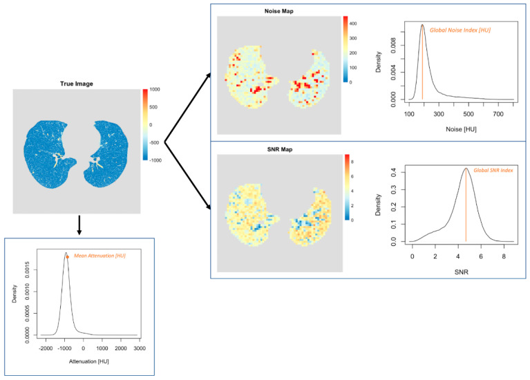
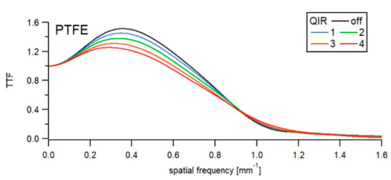
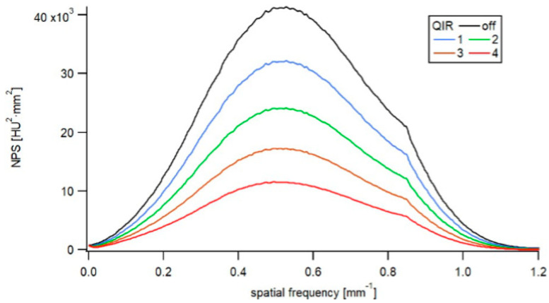
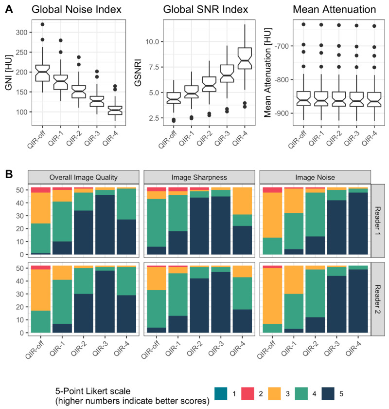
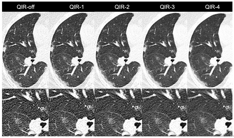
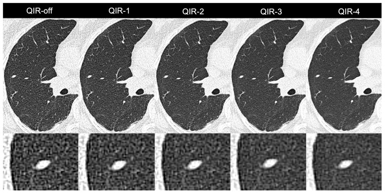
The aim of this study was to characterize image quality and to determine the optimal strength levels of a novel iterative reconstruction algorithm (quantum iterative reconstruction, QIR) for low-dose, ultra-high-resolution (UHR) photon-counting detector CT (PCD-CT) of the lung. Images were acquired on a clinical dual-source PCD-CT in the UHR mode and reconstructed with a sharp lung reconstruction kernel at different strength levels of QIR (QIR-1 to QIR-4) and without QIR (QIR-off). Noise power spectrum (NPS) and target transfer function (TTF) were analyzed in a cylindrical phantom. 52 consecutive patients referred for low-dose UHR chest PCD-CT were included (CTDIvol: 1 ± 0.6 mGy). Quantitative image quality analysis was performed computationally which included the calculation of the global noise index (GNI) and the global signal-to-noise ratio index (GSNRI). The mean attenuation of the lung parenchyma was measured. Two readers graded images qualitatively in terms of overall image quality, image sharpness, and subjective image noise using 5-point Likert scales. In the phantom, an increase in the QIR level slightly decreased spatial resolution and considerably decreased noise amplitude without affecting the frequency content. In patients, GNI decreased from QIR-off (202 ± 34 HU) to QIR-4 (106 ± 18 HU) (p < 0.001) by 48%. GSNRI increased from QIR-off (4.4 ± 0.8) to QIR-4 (8.2 ± 1.6) (p < 0.001) by 87%. Attenuation of lung parenchyma was highly comparable among reconstructions (QIR-off: -849 ± 53 HU to QIR-4: -853 ± 52 HU, p < 0.001). Subjective noise was best in QIR-4 (p < 0.001), while QIR-3 was best for sharpness and overall image quality (p < 0.001). Thus, our phantom and patient study indicates that QIR-3 provides the optimal iterative reconstruction level for low-dose, UHR PCD-CT of the lungs.
Keywords: X-ray computed; imaging; lung; phantoms; tomography.
Conflict of interest statement
T.F. is an employee of Siemens Healthineers. The remaining authors declare that the research was conducted in the absence of any commercial or financial relationships that could be construed as a potential conflict of interest.
Figures
Similar articles
-
Voxelwise characterization of noise for a clinical photon-counting CT scanner with a model-based iterative reconstruction algorithm.Eur Radiol Exp. 2025 Jan 2;9(1):2. doi: 10.1186/s41747-024-00541-2. Eur Radiol Exp. 2025. PMID: 39747757 Free PMC article.
-
Quantum Iterative Reconstruction for Abdominal Photon-counting Detector CT Improves Image Quality.Radiology. 2022 May;303(2):339-348. doi: 10.1148/radiol.211931. Epub 2022 Feb 1. Radiology. 2022. PMID: 35103540
-
Ultra-high resolution coronary CT angiography on photon-counting detector CT: bi-centre study on the impact of quantum iterative reconstruction on image quality and accuracy of stenosis measurements.Eur J Radiol. 2024 Jul;176:111517. doi: 10.1016/j.ejrad.2024.111517. Epub 2024 May 18. Eur J Radiol. 2024. PMID: 38805884
-
Reconstruction Kernel Optimization for Ultra-High-Resolution Photon-Counting Detector Computed Tomography of the Lung.J Comput Assist Tomogr. 2025 May-Jun 01;49(3):456-461. doi: 10.1097/RCT.0000000000001694. Epub 2024 Nov 18. J Comput Assist Tomogr. 2025. PMID: 39761487
-
Ultra-High-Resolution Photon-Counting CT Imaging of the Chest: A New Era for Morphology and Function.Invest Radiol. 2023 Jul 1;58(7):482-487. doi: 10.1097/RLI.0000000000000968. Epub 2023 Mar 11. Invest Radiol. 2023. PMID: 36897831 Review.
Cited by
-
[CT technology: photon-counting detector computed tomography].Radiologie (Heidelb). 2023 Jul;63(7):497-506. doi: 10.1007/s00117-023-01166-z. Epub 2023 Jun 8. Radiologie (Heidelb). 2023. PMID: 37289254 Free PMC article. Review. German.
-
Visibility of Intracranial Perforating Arteries Using Ultra-High-Resolution Photon-Counting Detector Computed Tomography (CT) Angiography.Tomography. 2024 Nov 21;10(12):1867-1880. doi: 10.3390/tomography10120136. Tomography. 2024. PMID: 39728898 Free PMC article.
-
Photon-counting detector CT: early clinical experience review.Br J Radiol. 2023 Jul;96(1147):20220544. doi: 10.1259/bjr.20220544. Epub 2023 Feb 10. Br J Radiol. 2023. PMID: 36744809 Free PMC article. Review.
-
Voxelwise characterization of noise for a clinical photon-counting CT scanner with a model-based iterative reconstruction algorithm.Eur Radiol Exp. 2025 Jan 2;9(1):2. doi: 10.1186/s41747-024-00541-2. Eur Radiol Exp. 2025. PMID: 39747757 Free PMC article.
-
Technology Characterization Through Diverse Evaluation Methodologies: Application to Thoracic Imaging in Photon-Counting Computed Tomography.J Comput Assist Tomogr. 2025 Jan-Feb 01;49(1):113-124. doi: 10.1097/RCT.0000000000001608. Epub 2024 Apr 15. J Comput Assist Tomogr. 2025. PMID: 38626754 Free PMC article.
References
-
- Sartoretti T., Eberhard M., Rüschoff J.H., Pietsch H., Jost G., Nowak T., Schmidt B., Flohr T., Euler A., Alkadhi H. Photon-counting CT with tungsten as contrast medium: Experimental evidence of vessel lumen and plaque visualization. Atherosclerosis. 2020;310:11–16. doi: 10.1016/j.atherosclerosis.2020.07.023. - DOI - PubMed
-
- Yu Z., Leng S., Li Z., Halaweish A.F., Kappler S., Ritman E.L., McCollough C.H. How Low Can We Go in Radiation Dose for the Data-Completion Scan on a Research Whole-Body Photon-Counting Computed Tomography System. J. Comput. Assist. Tomogr. 2016;40:663–670. doi: 10.1097/RCT.0000000000000412. - DOI - PMC - PubMed
-
- Nowak T., Eberhard M., Schmidt B., Frey D., Distler O., Saltybaeva N., Alkadhi H., Euler A. Bone Mineral Density Quantification from Localizer Radiographs: Accuracy and Precision of Energy-integrating Detector CT and Photon-counting Detector CT. Radiology. 2021;298:147–152. doi: 10.1148/radiol.2020202767. - DOI - PubMed
LinkOut - more resources
Full Text Sources
