. 2022 Feb 9;12(2):281.
doi: 10.3390/biom12020281.

Systemic Treatment with Pioglitazone Reverses Vision Loss in Preclinical Glaucoma Models

Affiliations

Systemic Treatment with Pioglitazone Reverses Vision Loss in Preclinical Glaucoma Models

Huilan Zeng et al. Biomolecules. .

Abstract

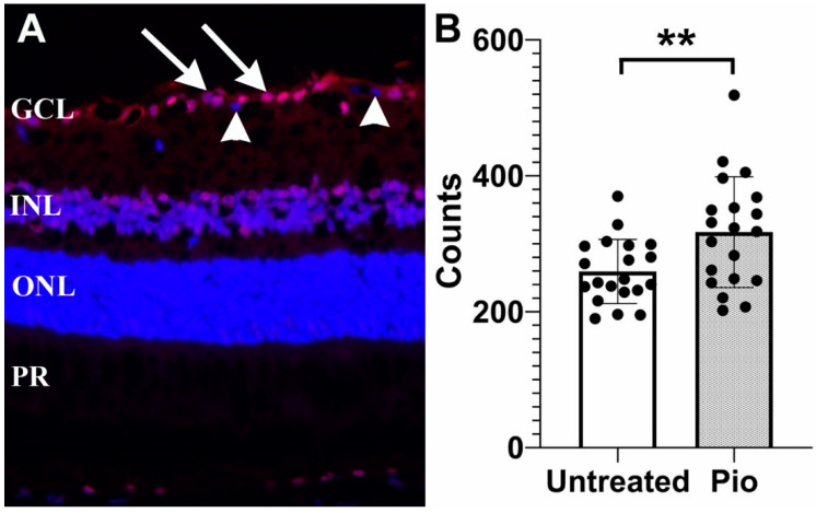
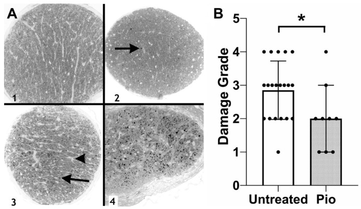
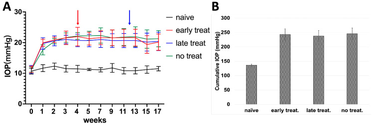
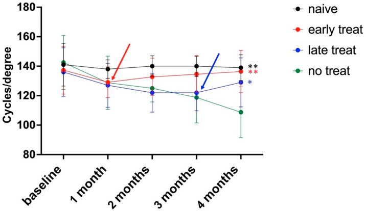
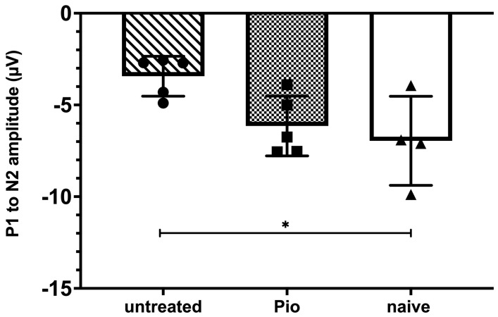
Neuroinflammation significantly contributes to the pathophysiology of several neurodegenerative diseases. This is also the case in glaucoma and may be a reason why many patients suffer from progressive vision loss despite maximal reduction in intraocular pressure. Pioglitazone is an agonist of the peroxisome proliferator-activated receptor gamma (PPARγ) whose pleiotrophic activities include modulation of cellular energy metabolism and reduction in inflammation. In this study we employed the DBA2/J mouse model of glaucoma with chronically elevated intraocular pressure to investigate whether oral low-dose pioglitazone treatment preserves retinal ganglion cell (RGC) survival. We then used an inducible glaucoma model in C57BL/6J mice to determine visual function, pattern electroretinographs, and tracking of optokinetic reflex. Our findings demonstrate that pioglitazone treatment does significantly protect RGCs and prevents axonal degeneration in the glaucomatous retina. Furthermore, treatment preserves and partially reverses vision loss in spite of continuously elevated intraocular pressure. These data suggest that pioglitazone may provide treatment benefits for those glaucoma patients experiencing continued vision loss.

Keywords: PPARγ; glaucoma; glucose metabolism; pioglitazone.


Conflict of interest statement

The authors declare no conflict of interest.

Figures

Figure 1
Figure 1
Protective effect of pioglitazone in DBA/2J mice. (A) Representative image showing γ-synuclein labeling of RGCs (arrows). Displaced amacrine cells are not bound by this antibody and are not counted (arrowheads) (B) RGC density in pioglitazone treated mice (Pio) is significantly higher than in untreated controls (p = 0.009). Each dot represents one eye. GCL: Ganglion cell layer; INL: Inner nuclear layer, ONL: Outer nuclear layer, PR: Photoreceptor inner segments. **: p < 0.01.
Figure 2
Figure 2
Optic nerve damage in DBA2/J mice. (A) Examples of optic nerve damage grades. A grade of 1 indicates a normal optic nerve, whereas a grade of 4 is assigned to optic nerves displaying severe damage and numerous PPD stained axons (arrows) and areas of gliosis (arrowhead). (B) Grading of optic nerve damage demonstrates a protective effect in pioglitazone (Pio)-treated mice (p = 0.0334). Each dot represents one optic nerve. *: p < 0.05.
Figure 3
Figure 3
IOP in C57BL/6J mice following injection of Ad5myoc into the anterior chamber. (A) IOP profile of treatment groups. Pioglitazone treatment was initiated either 4 weeks (red arrow) or 12 weeks (blue arrow) after induction of elevated IOP. (B) Average cumulative IOP in treatment groups. No significant differences in cumulative IOP were observed between Ad5myoc-injected mice (p > 0.25).
Figure 4
Figure 4
Pioglitazone improves visual acuity in glaucomatous mice. Induction of elevated IOP following baseline measurements leads to a decrease in OKR. Pioglitazone treatment after one month (early treatment, red line) caused a reversal of these deficits. Pioglitazone treatment in mice with more advanced damage (late treatment group, blue line) prevents further functional decline and partially reverses losses. Arrows indicate the start of pioglitazone treatments. ** p < 0.01; * p > 0.05.
Figure 5
Figure 5
Pattern electroretinography (pERG) after 8 weeks of elevated IOP. The P1 to N2 amplitude is significantly diminished in untreated mice when compared to naïve animals (p = 0.028). Animals receiving pioglitazone in the drinking water for 28 days before pERG measurements did not display significant differences when compared to naïve mice (p = 0.72). *: p < 0.05.

References

    1. Jonas J.B., Aung T., Bourne R.R., Bron A.M., Ritch R., Panda-Jonas S. Glaucoma. Lancet. 2017;390:2183–2193. doi: 10.1016/S0140-6736(17)31469-1. - DOI - PubMed
    1. Quigley H.A., Broman A.T. The number of people with glaucoma worldwide in 2010 and 2020. Br. J. Ophthalmol. 2006;90:262–267. doi: 10.1136/bjo.2005.081224. - DOI - PMC - PubMed
    1. Weinreb R.N., Khaw P.T. Primary open-angle glaucoma. Lancet. 2004;363:1711–1720. doi: 10.1016/S0140-6736(04)16257-0. - DOI - PubMed
    1. Gharahkhani P., Jorgenson E., Hysi P., Khawaja A.P., Pendergrass S., Han X., Ong J.S., Hewitt A.W., Segre A.V., Rouhana J.M., et al. Genome-wide meta-analysis identifies 127 open-angle glaucoma loci with consistent effect across ancestries. Nat. Commun. 2021;12:1258. doi: 10.1038/s41467-020-20851-4. - DOI - PMC - PubMed
    1. Flammer J., Orgul S., Costa V.P., Orzalesi N., Krieglstein G.K., Serra L.M., Renard J.P., Stefansson E. The impact of ocular blood flow in glaucoma. Prog. Retin. Eye Res. 2002;21:359–393. doi: 10.1016/S1350-9462(02)00008-3. - DOI - PubMed

Publication types