Review
. 2022 Feb 11;12(2):291.
doi: 10.3390/biom12020291.

JAK2 Variant Signaling: Genetic, Hematologic and Immune Implication in Chronic Myeloproliferative Neoplasms

Affiliations
Review

JAK2 Variant Signaling: Genetic, Hematologic and Immune Implication in Chronic Myeloproliferative Neoplasms

Dania G Torres et al. Biomolecules. .

Abstract

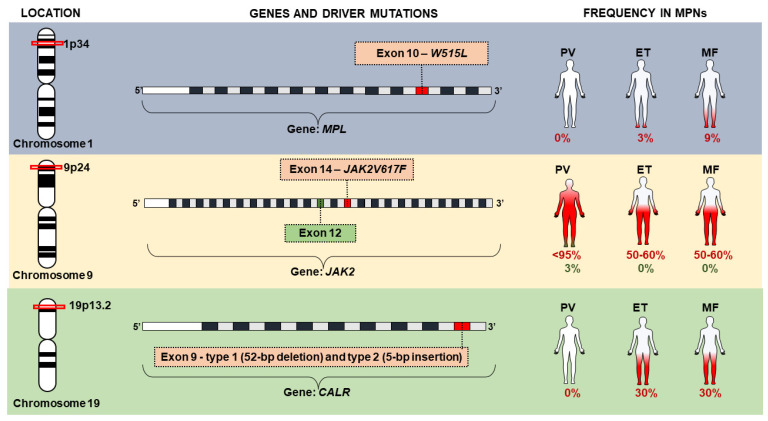
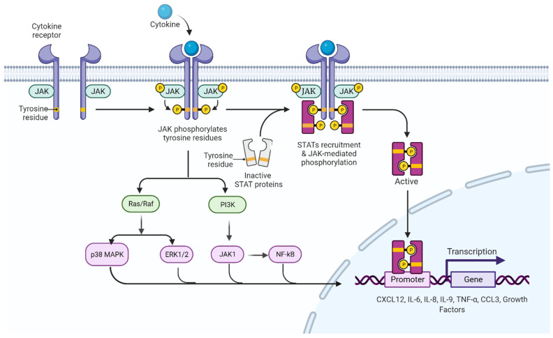
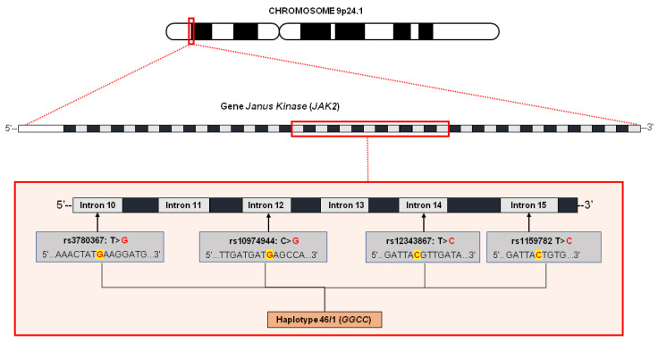
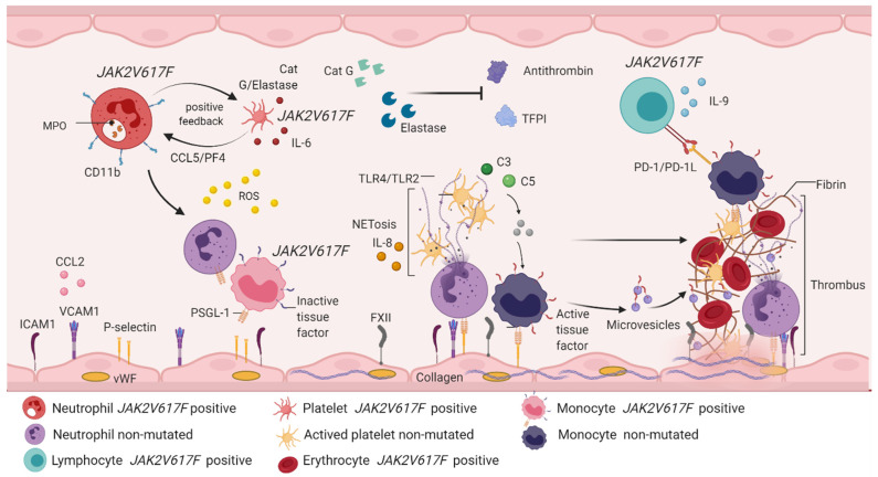
The JAK2V617F variant constitutes a genetic alteration of higher frequency in BCR/ABL1 negative chronic myeloproliferative neoplasms, which is caused by a substitution of a G ˃ T at position 1849 and results in the substitution of valine with phenylalanine at codon 617 of the polypeptide chain. Clinical, morphological and molecular genetic features define the diagnosis criteria of polycythemia vera, essential thrombocythemia and primary myelofibrosis. Currently, JAK2V617F is associated with clonal hematopoiesis, genomic instability, dysregulations in hemostasis and immune response. JAK2V617F clones induce an inflammatory immune response and lead to a process of immunothrombosis. Recent research has shown great interest in trying to understand the mechanisms associated with JAK2V617F signaling and activation of cellular and molecular responses that progressively contribute to the development of inflammatory and vascular conditions in association with chronic myeloproliferative neoplasms. Thus, the aim of this review is to describe the main genetic, hematological and immunological findings that are linked to JAK2 variant signaling in chronic myeloproliferative neoplasms.

Keywords: JAK2V617F signaling; clonal hematopoiesis; hemostasis; immune response; immunothrombosis; myeloproliferative neoplasms.


Conflict of interest statement

The authors declare no conflict of interest.

Figures

Figure 1
Figure 1
Timeline of myeloproliferative neoplasms. In gray, the clinical era of the MPNs can be seen, which is based on semiological aspects. In orange, the genetic era of MPNs is highlighted, since associated genetic research made it possible to identify genetic alterations that are markers of some MPNs, an important factor that supports diagnosis. CML: chronic myeloid leukemia; BCR/ABL: BCR/ABL genetic rearrangement.
Figure 2
Figure 2
Driver mutations in JAK2, MPL and CALR genes associated with chronic myeloproliferative neoplasms (MPNs).
Figure 3
Figure 3
JAK/STAT signaling pathway. In the absence of cytokines, the JAK protein remains inactive in regions close to the intracellular domains of the receptor. When a cytokine binds to a receptor, JAK proteins and intracellular domains of the receptor are phosphorylated, activating and recruiting STAT proteins, which dimerize and translocate to the nucleus to initiate the transcription process of genes involved in cell proliferation. In addition to the activation of the JAK/STAT pathway, there is an interconnection with other intracellular signaling pathways, among the most prominent are the Ras/Raf/MAPK pathway and the PI3K pathway; the latter manifesting interconnection with JAK1 proteins, which indirectly activate the NFkB pathway, the transcriptional factors that activate the production of cytokines (among them CXCL12, IL-6, IL-8, IL-9, TNF-α and CCL3) and the growth factors identified in the inflammatory profile of individuals with chronic myeloproliferative neoplasms. JAK: Janus kinase protein; STAT: signal transducers and activators of transcription; MAPK: mitogen-activated protein kinase; PI3K: phosphoinositol kinase 3; JAK1: Janus kinase class 1 protein; NFkB: light chain nuclear factor B-cell kappa; CXCL12: chemokine 12 with CXC motif; IL-6: interleukin 6; IL-8: interleukin 8; IL-9: interleukin 9; TNF-α: tumor necrosis factor alpha; CCL3: chemokine ligand 3.
Figure 4
Figure 4
Characterization of the 46/1 haplotype without JAK2 gene. The haplotype, also called GGCC, is altered by four variants located in intron 10 (rs3780367: T > G), intron 12 (rs10974944: C > G), intron 14 (rs12343867: T > C) and intron 15 (rs1159782: T > C). It is believed that the presence of this haplotype conditions an increase in the mutation rate of the gene locus in question, thus resulting in the emergence of mutations with a selective advantage, as in the case of JAK2V617F.
Figure 5
Figure 5
Immunothrombosis involves the participation of hematopoietic cells and immune system cells. JAK2V617F positive neutrophils express CD11b on the cell surface and secrete enzymes such as MPO, cathepsin G and elastase. The release of cathepsin G and elastase activates CCL5/PF4 signaling in JAK2V617F positive platelets, which is an interaction that creates positive feedback between platelets and neutrophils, and favors the production of IL-6 and ROS. Free cathepsin and elastase inhibit the function of antithrombin and plasma TFPI, contributing to the thrombotic phenotype. Unmutated neutrophils are activated by the action of ROS, which recruit and activate monocytes with PSGL-1 expression and inactive tissue factor. Both cells manifest rolling and endothelial adhesion by binding adhesion molecules expressed in endothelial cells (ICAM1, VCAM1, P-selectin, CCL2 and FXII). Simultaneously, endothelial cells are activated by the action of ROS, releasing vWF and collagen, thus forming a platelet–monocyte–neutrophil interaction, which favors the discharge of genetic and protein material from the neutrophil, starting with NETosis. This brings with it the expression of IL-8 and activation of TLR4/TLR2 in platelets, a fact that allows the expression of receptor glycoproteins and, therefore, platelet aggregation. In NETosis, erythrocytes are recruited and release hemoglobin and increase ROS production. The complement system is active in response to activation of immune cells, especially through the action of C3 and C5, which induces signaling from JAK2V617F positive monocytes and non-mutated monocytes. JAK2V617F positive monocytes express PD-1L, which immediately activates PD-1 in JAK2V617F lymphocytes, a mechanism responsible for the evasion of the immune response described in chronic myeloproliferative neoplasms. Recruitment and activation of immune and hematopoietic cells benefits the conversion of fibrinogen into fibrin, giving rise to thrombus formation, the main factor involved in vascular complications described in myeloproliferative neoplasms. MPO: myeloperoxidase; CCL5: chemokine ligand 5; PF4: platelet factor 4; IL-6: interleukin 6; ROS: reactive oxygen species; TFPI: plasma tissue factor inhibitor; PSGL-1: P-selectin ligand 1; ICAM-1: intercellular adhesion molecules 1; VCAM-1: vascular cell adhesion molecule 1; CCL2: chemokine ligand 2; FXII: factor XII; vWF: Von Willebrand factor; IL-8: interleukin 8; TLR2/TLR4: Toll-like receptor 2/4; C3: complement component 3; C5: complement component 5; PD-1: programmed death protein; PD-L1: programmed death protein ligand 1.

References

    1. Tremblay D., Yacoub A., Hoffman R. Overview of Myeloproliferative Neoplasms: History, Pathogenesis, Diagnostic Criteria, and Complications. Hematol. Oncol. Clin. N. Am. 2021;35:159–176. doi: 10.1016/j.hoc.2020.12.001. - DOI - PMC - PubMed
    1. Chauffaille M. Neoplasias mieloproliferativas: Revisão dos critérios diagnósticos e dos aspectos clínicos. Rev. Bras. Hematol. Hemoter. 2010;32:308–316. doi: 10.1590/S1516-84842010005000091. - DOI
    1. Nangalia J., Green A.R. Myeloproliferative neoplasms: From origins to outcomes. Hematology. 2017;1:470–479. doi: 10.1182/asheducation-2017.1.470. - DOI - PMC - PubMed
    1. Tefferi A. The history of myeloproliferative disorders: Before and after Dameshek. Leukemia. 2008;22:3–13. doi: 10.1038/sj.leu.2404946. - DOI - PubMed
    1. Tefferi A. Myeloproliferative neoplasms: A decade of discoveries and treatment advances. Am. J. Hematol. 2016;91:50–58. doi: 10.1002/ajh.24221. - DOI - PubMed

MeSH terms

LinkOut - more resources