. 2022 Jan 22;13(2):193.
doi: 10.3390/genes13020193.

MtWRP1, a Novel Fabacean Specific Gene, Regulates Root Nodulation and Plant Growth in Medicago truncatula

Affiliations

MtWRP1, a Novel Fabacean Specific Gene, Regulates Root Nodulation and Plant Growth in Medicago truncatula

Wei Chen et al. Genes (Basel). .

Abstract

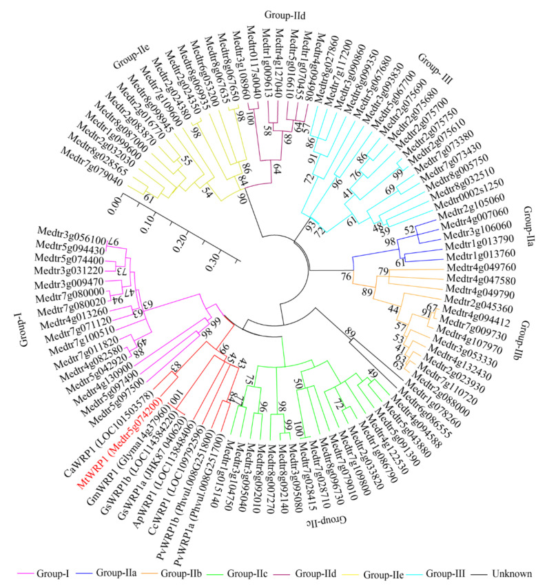
Fabaceans symbiotically interact with nitrogen-fixing rhizobacteria to form root nodules. Some fabacean specific proteins play important roles in the symbiosis. WRKY-related Protein (WRP) is a novel fabacean specific protein, whose functions have not been well characterized. In this study, MtWRP1 was functionally characterized in Medicago truncatula. It contains a WRKY domain at C-terminal and a novel transmembrane (TM) domain at N-terminal, and its WRKY domain was highly similar to the N-terminal WRKY domain of the group I WRKY proteins. The TM domain was highly homologous to the eukaryotic cytochrome b561 (Cytb561) proteins from birds. Subcellular localization revealed that MtWRP1 was targeted to the Golgi apparatus through the novel TM domain. MtWRP1 was highly expressed in roots and nodules, suggesting its possible roles in the regulation of root growth and nodulation. Both MtWRP1-overexpression transgenic M. truncatula and MtWRP1 mutants showed altered root nodulation and plant growth performance. Specifically, the formation of root nodules was significantly reduced in the absence of MtWRP1. These results demonstrated that MtWRP1 plays critical roles in root nodulation and plant growth.

Keywords: Medicago truncatula; MtWRP1; N-terminal transmembrane domain; nodulation; plant growth.


Conflict of interest statement

The authors declare no conflict of interest.

Figures

Figure 1
Figure 1
Multiple amino acid sequence alignment: (A) Close homologues of MtWRP1 in fabaceans. Amino acid sequences of the WRPs from M. truncatula (MtWRP1), Cicer arietinum (CaWRP1), Abrus precatorius (ApWRP1), Cajanus cajan (CcWRP1), Glycine soja (GsWRP1a and GsWRP1b), Glycine max (GmWRP1), and Phaseolus vulgaris (PvWRP1a and PvWRP1b). The TM domains of WRPs were indicated by blue lines. The WRKY domain of WRPs were indicated by red lines. The putative localization signals were indicated by the green box. (B) Alignment of the MtWRP1 WRKY domain sequence with the consensus sequences of the WRKY domains of seven WRKY subfamilies, including the N-terminal (NT) and C-terminal (CT) WRKY domains of group I WRKY proteins. The core sequences of the WRKY domain were indicated by triangles.
Figure 2
Figure 2
Structural analysis of TM domain at N-terminal MtWRP1: (A) Prediction of conserved TM domain in MtWRP1 as 5-passage transmembrane domain. (B) Sequence comparison of the TM domains of MtWRP1, FAcytb561 (XP_005049393.1), and TGcytb561 (XP_002198627.1).
Figure 3
Figure 3
The phylogenetic analysis of MtWRP1, WRPs in different fabacean plants and the WRKYs in M. truncatula. The phylogenetic tree was generated from full-length amino acid sequences by MEGA 5 using the Neighbor-joining method. Numbers on branches indicated bootstrap values for 1000 replicates. The scale bar indicated the length of branch, reflecting the ancient divergence of these genes from each other.
Figure 4
Figure 4
Subcellular localization: (A) Subcellular localization of MtWRP1 and TM domain at N-terminal of MtWRP1 (MtWRP1-NTD). A majority of MtWRP1-GFP and MtWRP1-NTD-GFP fluorescence signals were overlapped with mRFP Golgi marker signals. The arrows indicated the localization of GFP-fused proteins overlapped with the mRFP Golgi marker signals. Co-localization of free GFPs driven by CaMV 35S and mRFP Golgi marker were used as control. (B) Subcellular localization of WRKY domain at C-terminal of MtWRP1 (MtWRP1-CTD). MtWRP1-CTD-GFP fluorescence signals were overlapped with mRFP nucleus marker signals. Co-localization of free GFPs and mRFP nucleus marker were used as control. Bar, 20 µm.
Figure 5
Figure 5
Expression of MtWRP1 in M. truncatula: (A) The transcript levels of MtWRP1 in different plant organs. Young leaves, stems, and roots were collected from 4-week-old plants. Flowers were sampled from 8-week-old plants. Pods at pod-bearing period and seeds at seed-filling period were collected from 90-day-old plants. (B) The transcript levels of MtWRP1 in nodules (Nod 15, 20, and 30 dpi) inoculated with S. mliloti 1021. The gene expression in root at 15 dpi (Root-15 dpi) was used as control. Data represent mean ± SD (n = three independent biological replicates). Different letters above the standard error bars indicated a significant difference between the simples as determined by Duncan’s multiple range test (p < 0.05).
Figure 6
Figure 6
Identification of overexpression transgenic lines and MtWRP1 mutants in M. truncatula: (A) The transcript levels of MtWRP1 in transgenic M. truncatula. 1–10: transgenic lines. Data represent mean ± SD (n = three independent biological replicates). (B) Schematic representation of MtWRP1 gene model and the Tnt1 insertion sites. The box indicated the coding region of MtWRP1, which contained only one exon. The position of Tnt1 insertions was indicated in base pairs (bp) with arrowheads. The orientation of Tnt1 insertions was indicated by black arrows. (C) RT-PCR analysis of MtWRP1 expression in wrp1-1. Lane 1-1, 2-1, and R108-1 were PCR-based identification of Tnt1 insertion (544 bp) using primers MtWRP1-R and Tnt1-F2. Lane 1-2, 2-2 and R108-2 were PCR-based identification of MtWRP1 (984 bp) using primers MtWRP1-F and MtWRP1-R. R108 was used as the wild type control. M was a DL2000 DNA marker. (D) RT-PCR analysis of MtWRP1 expression in wrp1-2. Lane 1-1, 2-1, and R108-1 were PCR-based identification of Tnt1 insertion (677 bp) using primers MtWRP1-F and Tnt1-F2. Lane 1-2, 2-2, and R108-2 were PCR-based identification of MtWRP1 (984 bp) using primers MtWRP1-F and MtWRP1-R. R108 was control. M was a DL2000 DNA marker. Data represent mean ± SD (n = three independent biological replicates). Different letters above the standard error bars indicated a significant difference between the lines as determined by Duncan’s multiple range test (p < 0.05).
Figure 7
Figure 7
Effects of MtWRP1 on plant growth and development in M. truncatula: (A) Growth phenotype of MtWRP1 mutants (wrp1-1 and wrp1-2), R108 and transgenic M. truncatula (OE1, OE2, and OE3) at 30 days post inoculation with S. mliloti 1021 under nitrogen-deficient conditions. (B) The plant heights of MtWRP1 mutants, R108, and transgenic M. truncatula. (C) The shoot fresh weights of MtWRP1 mutants, R108, and transgenic M. truncatula. Data represent mean ± SD (n = four independent biological replicates). Different letters above the standard error bars indicated a significant difference between the lines as determined by Duncan’s multiple range test (p < 0.05).
Figure 8
Figure 8
Effects of MtWRP1 on root growth and nodulation in M. truncatula: (A) Nodulation phenotype of MtWRP1 mutants (wrp1-1 and wrp1-2), R108, and transgenic M. truncatula (OE1, OE2, and OE3) at 30 dpi under nitrogen-deficient conditions. Arrowheads indicated nodules. Bar, 1 cm. (B) The root fresh weights of MtWRP1 mutants, R108, and transgenic M. truncatula. (C) The root total lengths of MtWRP1 mutants, R108, and transgenic M. truncatula. (D) The root tips of MtWRP1 mutants, R108 and transgenic M. truncatula. (E) The nodule number of MtWRP1 mutants, R108, and transgenic M. truncatula. (F) The nodule weights of MtWRP1 mutants, R108, and transgenic M. truncatula. Data represent mean ± SD (n = four independent biological replicates). Different letters above the standard error bars indicated a significant difference between the lines as determined by Duncan’s multiple range test (p < 0.05).

References

    1. Wang J., Andersen S.U., Ratet P. Molecular and Cellular Mechanisms of the Legume-Rhizobia Symbiosis. Front. Plant Sci. 2018;9:1839. doi: 10.3389/fpls.2018.01839. - DOI - PMC - PubMed
    1. Peix A., Ramírez-Bahena M.H., Velázquez E., Bedmar E.J. Bacterial Associations with Legumes. Crit. Rev. Plant Sci. 2014;34:17–42. doi: 10.1080/07352689.2014.897899. - DOI
    1. Peoples M.B., Ladha D.F., Herridge J.K. Biological Nitrogen Fixation: An Efficient Source of Nitrogen for Sustainable Agricultural Production? Plant Soil. 1995;174:3–28. doi: 10.1007/BF00032239. - DOI
    1. Koskey G., Mburu S.W., Njeru E.M., Kimiti J.M., Ombori O., Maingi J.M. Potential of Native Rhizobia in Enhancing Nitrogen Fixation and Yields of Climbing Beans (Phaseolus vulgaris L.) in Contrasting Environments of Eastern Kenya. Front. Plant Sci. 2017;8:443. doi: 10.3389/fpls.2017.00443. - DOI - PMC - PubMed
    1. Oldroyd G.E., Downie J.A. Coordinating Nodule Morphogenesis with Rhizobial Infection in Legumes. Annu. Rev. Plant Biol. 2008;59:519–546. doi: 10.1146/annurev.arplant.59.032607.092839. - DOI - PubMed

Publication types

LinkOut - more resources