. 2022 Feb 16;13(2):357.
doi: 10.3390/genes13020357.

A Split NanoLuc Reporter Quantitatively Measures Circular RNA IRES Translation

Affiliations

A Split NanoLuc Reporter Quantitatively Measures Circular RNA IRES Translation

Priyanka Sehta et al. Genes (Basel). .

Abstract

Internal ribosomal entry sites (IRESs) are RNA secondary structures that mediate translation independent from the m7G RNA cap. The dicistronic luciferase assay is the most frequently used method to measure IRES-mediated translation. While this assay is quantitative, it requires numerous controls and can be time-consuming. Circular RNAs generated by splinted ligation have been shown to also accurately report on IRES-mediated translation, however suffer from low yield and other challenges. More recently, cellular sequences were shown to facilitate RNA circle formation through backsplicing. Here, we used a previously published backsplicing circular RNA split GFP reporter to create a highly sensitive and quantitative split nanoluciferase (NanoLuc) reporter. We show that NanoLuc expression requires backsplicing and correct orientation of a bona fide IRES. In response to cell stress, IRES-directed NanoLuc expression remained stable or increased while a capped control reporter decreased in translation. In addition, we detected NanoLuc expression from putative cellular IRESs and the Zika virus 5' untranslated region that is proposed to harbor IRES function. These data together show that our IRES reporter construct can be used to verify, identify and quantify the ability of sequences to mediate IRES-translation within a circular RNA.

Keywords: RNA circles; internal ribosome entry site; reporter assay; split NanoLuc.


Conflict of interest statement

The authors declare that they have no conflict of interest with the contents of this article. The content is solely the responsibility of the authors.

Figures

Figure 1
Figure 1
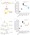
A split NanoLuc reporter quantitatively measures translation from a circular RNA. (A) ZKSCAN split NanoLuc plasmid construct contains SbfI and EcoRV restriction sites, into which the EMCV IRES had been inserted. Upon transcription, a linear capped and polyadenylated transcript is formed. Upon backsplicing, the split NanoLuc open reading frame (ORF) is fused, which results in active NanoLuc protein. (B) NanoLuc over firefly luciferase luminescence ratios is plotted for the different control constructs containing the EMCV IRES in the forward (EMCV) or reverse orientation (EMCV Rev), the EMCV IRES in the forward direction with the intronic splice donor (∆ splice donor) or acceptor sites (∆ splice acceptor) deleted, co-expression of N− and C−terminal fragments (N + C halves) from two co-transfected DNA plasmids, and the empty plasmid with a short 9 bp unstructured linker inserted into the SbfI and EcoRV sites (linker). Schematics corresponding to the constructs tested here are shown to the right. (C) NanoLuc expression from circular RNAs in response to cellular stress induced by sodium arsenite, tunicamycin and thapsigargin compared to an untreated and a DMSO control. NanoLuc expression normalized to firefly luciferase is displayed in a percentage. All experiments were performed at least in triplicate. Error bars represent the standard deviation. Significances were calculated using an unpaired t test with Welch’s correction with p ≤ 0.05 = *, p ≤ 0.01 = **.
Figure 2
Figure 2
The relative activity of cellular IRESs can be quantified with a split NanoLuc reporter. (A) Putative cellular IRES sequences for c-Myc, DAP5 and c-Jun were cloned and inserted into the SbfI and EcoRV sites of the split NanoLuc reporter, generating a linear and a circular RNA. (B) Relative activity of cellular IRESs is measured by NanoLuc over firefly luciferase ratio. All experiments were performed at least in triplicate. Error bars represent the standard deviation. (C) ZIKV 5′ and 3′ UTRs RNA secondary structures and secondary structures in a combination of 5′-3′, 3′-5′ UTRs (D) NanoLuc over firefly luciferase ratio from ZIKV 5ʹ and 3ʹ UTRs alone, or in a combination of 5′-3′, 3′-5′ UTRs. Data for co-expression of N− and C−terminal fragments (N + C halves), the empty plasmid with a short 9 bp unstructured linker, and the EMCV IRES are identical to the data from Figure 1B, however, were plotted again for direct comparison. All experiments were performed at least in triplicate. Error bars represent the standard deviation. Significances were calculated using an unpaired t test with Welch’s correction with p ≤ 0.05 = *, p ≤ 0.01 = **.
Figure 3
Figure 3
A split NanoLuc reporter plasmid with a firefly luciferase reporter as an internal control. (A) Schematic of the split NanoLuc reporter plasmid with the firefly luciferase inserted into the MCS of pcDN3.1. Upon transcription, the linear RNA contains both the firefly luciferase open reading sequence and the split NanoLuc ORF. The linear RNA yields active firefly luciferase protein, while the split NanoLuc protein remains inactive. Upon backsplicing, the NanoLuc ORF remains in the circular RNA, is fused and yields NanoLuc protein. (B) NanoLuc over firefly luciferase values is plotted for the linker control and the c-Myc cellular IRES. Experiments were performed in triplicate. Error bars represent the standard deviation. Significances were calculated using an unpaired t test with Welch’s correction with p ≤ 0.001 = ***.

References

    1. Ramanathan A., Robb G.B., Chan S.-H. mRNA capping: Biological functions and applications. Nucleic Acids Res. 2016;44:7511–7526. doi: 10.1093/nar/gkw551. - DOI - PMC - PubMed
    1. Sonenberg N., Hinnebusch A.G. Regulation of translation initiation in eukaryotes: Mechanisms and biological targets. Cell. 2009;136:731–745. doi: 10.1016/j.cell.2009.01.042. - DOI - PMC - PubMed
    1. Merrick W.C., Pavitt G.D. Protein Synthesis Initiation in Eukaryotic Cells. Cold Spring Harb. Perspect. Biol. 2018;10:a033092. doi: 10.1101/cshperspect.a033092. - DOI - PMC - PubMed
    1. Pelletier J., Graff J., Ruggero D., Sonenberg N. Targeting the eIF4F translation initiation complex: A critical nexus for cancer development. Cancer Res. 2015;75:250–263. doi: 10.1158/0008-5472.CAN-14-2789. - DOI - PMC - PubMed
    1. Robichaud N., Sonenberg N., Ruggero D., Schneider R.J. Translational Control in Cancer. Cold Spring Harb. Perspect. Biol. 2018;11:a032896. doi: 10.1101/cshperspect.a032896. - DOI - PMC - PubMed

Publication types

MeSH terms

Substances