. 2022 Feb 16;13(2):359.
doi: 10.3390/genes13020359.

Maternal Uniparental Isodisomy of Chromosome 4 and 8 in Patients with Retinal Dystrophy: SRD5A3-Congenital Disorders of Glycosylation and RP1-Related Retinitis Pigmentosa

Affiliations

Maternal Uniparental Isodisomy of Chromosome 4 and 8 in Patients with Retinal Dystrophy: SRD5A3-Congenital Disorders of Glycosylation and RP1-Related Retinitis Pigmentosa

Nobutaka Tachibana et al. Genes (Basel). .

Abstract

Purpose: Uniparental disomy (UPD) is a rare chromosomal abnormality. We performed whole-exosome sequencing (WES) in cases of early-onset retinal dystrophy and identified two cases likely caused by UPD. Herein, we report these two cases and attempt to clarify the clinical picture of retinal dystrophies caused by UPD.

Methods: WES analysis was performed for two patients and their parents, who were not consanguineous. Functional analysis was performed in cases suspected of congenital disorders of glycosylation (CDG). We obtained clinical case data and reviewed the literature.

Results: In case 1, a novel c.57G>C, p.(Trp19Cys) variant in SRD5A3 was detected homozygously. Genetic analysis suggested a maternal UPD on chromosome 4, and functional analysis confirmed CDG. Clinical findings showed early-onset retinal dystrophy, intellectual disability, and epilepsy. In case 2, an Alu insertion (c.4052_4053ins328, p.[Tyr1352Alafs]) in RP1 was detected homozygously. Maternal UPD on chromosome 8 was suspected. The clinical picture was consistent with RP1-related retinitis pigmentosa. Although the clinical features of retinal dystrophy by UPD may vary, most cases present with childhood onset.

Conclusions: There have been limited reports of retinal dystrophy caused by UPD, suggesting that it is rare. Genetic counseling may be encouraged in pediatric cases of retinal dystrophy.

Keywords: RP1-related retinitis pigmentosa; SRD5A3 gene; congenital disorders of glycosylation; retinal dystrophy; uniparental isodisomy.


Conflict of interest statement

The authors declare no conflict of interest. The funders had no role in the design of the study; in the collection, analyses, or interpretation of data; in the writing of the manuscript, or in the decision to publish the results.

Figures

Figure 1
Figure 1
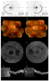
Clinical features of case 1. The visual field examined at the age of 9 years: (A) left eye; (B) right eye. Visual field examined at the age of 14 years: (C) left eye; (D) right eye. The visual field was progressively constricted. Fundus photograph obtained at the age of 9 years: (E) right eye; (F) left eye. Fundus photography showed poor retinal color and narrowing of the retinal blood vessels. OCT images obtained at the age of 10 years: (G) right eye; (H) left eye. Although OCT images were poor due to nystagmus, retinal thinning and EZ line disappearance were observed.
Figure 2
Figure 2
Findings of genetic and glycosylation analyses in patient 1. (A) (Left) H3M2 analysis using WES data showed LOH stretches on the whole of chromosome 4. (Right) Gene name, physical position, allele frequency in 8.3KJPN, and genotypes of the patient and her parents with 10 candidate variants are shown. Homozygous for reference allele, homozygous for alternate allele, and heterozygous genotypes are highlighted in yellow, green, and pink, respectively. Trio genotypes demonstrated maternal uniparental isodisomy. (B) Locations of reported SRD5A3 variants. Human SRD5A3 is 318 aa in length (NP_078868.1) and contains six putative transmembrane domains according to UniProt (Q9H8P0). Nonsense and frameshift variants are shown in the upper section, and missense variants are shown in the lower section. The p.Trp19Cys variant was in the transmembrane domain and substituted a conserved amino acid among vertebrates. (C) Deconvoluted spectra of transferrin from a patient (upper) and healthy individual (lower), analyzed by ESI-MS. The presence of the molecules lacking one or two N-glycans indicates deficient N-glycosylation.
Figure 3
Figure 3
Clinical features of case 2. The visual field examined at the age of 26 years: (A) left eye; (B) right eye. The visual field was constricted. Fundus photograph obtained at the age of 26 years: (C) right eye; (D) left eye. Fundus photography showed poor retinal color and narrowing of the retinal blood vessels. FAF obtained at the age of 31 years: (E) right eye; (F) left eye. FAF showed low fluorescence, consistent with retinal degeneration. OCT images obtained at the age of 31 years: (G) right eye; (H) left eye. OCT images showed that the retina was highly thinning, and the EZ line could not be observed at all.
Figure 4
Figure 4
Genetic findings in patient 2. (A) (Left) H3M2 analysis using WES data showed LOH stretches on the whole of chromosome 8. (Right) Gene name, physical position, allele frequency in 8.3KJPN, and genotypes of trio samples of eight candidate variants are shown. Homozygous for reference allele, homozygous for alternate allele, and heterozygous genotypes are highlighted in yellow, green, and pink, respectively. Trio genotypes indicated maternal uniparental isodisomy. (B) Integrative genomics viewer showed a sharp decline in sequencing depth at exon 4 of RP1 (red arrows), suggesting the Alu insertion. Discordant reads are highlighted in colors. (C) PCR analysis confirmed homozygous Alu insertion in the patient, and his mother was a carrier of the insertion.

References

    1. Engel E. A New Genetic Concept: Uniparental Disomy and Its Potential Effect, Isodisomy. Am. J. Med. Genet. 1980;6:137–143. doi: 10.1002/ajmg.1320060207. - DOI - PubMed
    1. Liehr T. Cytogenetic Contribution to Uniparental Disomy (UPD) Mol. Cytogenet. 2010;3:8. doi: 10.1186/1755-8166-3-8. - DOI - PMC - PubMed
    1. Spence J.E., Perciaccante R.G., Greig G.M., Willard H.F., Ledbetter D.H., Hejtmancik J.F., Pollack M.S., O’Brien W.E., Beaudet A.L. Uniparental Disomy as a Mechanism for Human Genetic Disease. Am. J. Hum. Genet. 1988;42:217–226. - PMC - PubMed
    1. Eggermann T. Prenatal Detection of Uniparental Disomies (UPD): Intended and Incidental Finding in the Era of Next Generation Genomics. Genes. 2020;11:1454. doi: 10.3390/genes11121454. - DOI - PMC - PubMed
    1. Mutirangura A., Greenberg F., Butler M.G., Malcolm S., Nicholls R.D., Chakravarti A., Ledbetter D.H. Multiplex PCR of Three Dinucleotide Repeats in the Prader-Willi/Angelman Critical Region (15q11-Q13): Molecular Diagnosis and Mechanism of Uniparental Disomy. Hum. Mol. Genet. 1993;2:143–151. doi: 10.1093/hmg/2.2.143. - DOI - PMC - PubMed

Publication types

MeSH terms

Substances

LinkOut - more resources