

Global Distribution and Clinical Features of Pythiosis in Humans and Animals
- PMID: 35205934
- PMCID: PMC8879638
- DOI: 10.3390/jof8020182
Global Distribution and Clinical Features of Pythiosis in Humans and Animals
Abstract
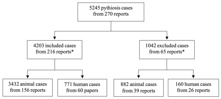
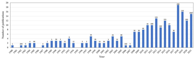
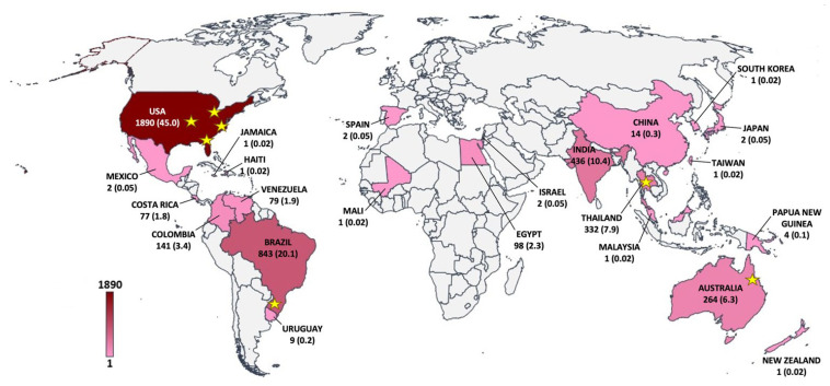
Pythiosis is a difficult-to-treat infectious disease caused by Pythium insidiosum. The condition is unfamiliar among healthcare workers. Manifestation of pythiosis is similar to other fungal infections, leading to misdiagnosis and delayed treatment. The geographical extent of pythiosis at a global scale is unclear. This study aimed to analyze the clinical information recorded in the scientific literature to comprehensively project epidemiological characteristics, clinical features, and future trends of pythiosis. From 1980 to 2021, 4203 cases of pythiosis in humans (n = 771; 18.3%) and animals (primarily horse, dog, and cow; n = 3432; 81.7%), with an average of 103 cases/year, were recruited. Pythiosis case reports significantly increased in the last decade. Pythiosis spanned 23 tropical, subtropical, and temperate countries worldwide. Some patients acquired pythiosis from a trip to an endemic country. Strikingly, 94.3% of human cases were in India and Thailand, while 79.2% of affected animals were in the U.S.A. and Brazil. Clinical features of pythiosis varied. Vascular and ocular pythiosis were only observed in humans, whereas cutaneous/subcutaneous and gastrointestinal infections were predominant in animals. Mortality depended on host species and clinical forms: for example, none in patients with ocular pythiosis, 0.7% in cows with a cutaneous lesion, 26.8% in humans with vascular disease, 86.4% in dogs with gastrointestinal pathology, and 100% in several animals with disseminated infection. In summary, this study reports up-to-date epidemiological and clinical features of pythiosis in humans and animals. It increases awareness of this life-threatening disease, as the illness or outbreak can exist in any country, not limited to the endemic areas.
Keywords: Pythium insidiosum; clinical feature; distribution; epidemiology; pythiosis.
Conflict of interest statement
The authors declare no conflict of interest.
Figures
References
-
- Guedes R., Zica K., Nogueira R. Ficomicose e Habronemose Cutânea. Estudo Retrospectivo de Casos Diagnosticados No Período de 1979 a 1996. Arq. Bras. Med. Vet. Zootec. 1998;50:465–468.
Grants and funding
LinkOut - more resources
Full Text Sources
