

Symptomatic Young Adults with ST-Segment Elevation-Acute Coronary Syndrome or Myocarditis: The Three-Factor Diagnostic Model
- PMID: 35207189
- PMCID: PMC8877187
- DOI: 10.3390/jcm11040916
Symptomatic Young Adults with ST-Segment Elevation-Acute Coronary Syndrome or Myocarditis: The Three-Factor Diagnostic Model
Abstract
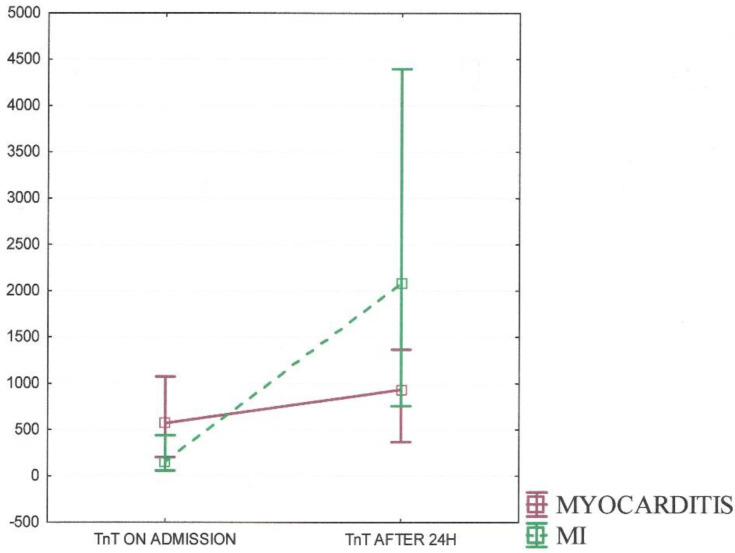
Myocarditis may mimic myocardial infarction (MI) due to a similar clinical presentation, including chest pain, electrocardiography changes, and laboratory findings. The purpose of the study was to investigate the diagnostic value of clinical, laboratory, and electrocardiography characteristics of patients with acute coronary syndrome - like myocarditis and MI. We analysed 90 patients (≤45 years old) with an initial diagnosis of ST-segment elevation myocardial infarction; 40 patients (44.4%), through the use of cardiac magnetic resonance, were confirmed to have myocarditis, and 50 patients (55.6%) were diagnosed with MI. Patients with myocarditis were younger and had fewer cardiovascular risk factors than those with MI. The cutoff value distinguishing between myocarditis and MI was defined as the age of 36 years. The history of recent infections (82.5% vs. 6%) and C-reactive protein (CRP) levels on admission (Me 45.9 vs. 3.4) was more associated with myocarditis. Further, the QTc interval was inversely correlated with the echocardiographic ejection fraction in both groups but was significantly longer in patients with MI. Non-invasive diagnostics based on clinical features and laboratory findings are basic but still essential tools for differentiation between MI and myocarditis. The three-factor model including the criteria of age, abnormal CRP, and history of recent infections might become a valuable clinical indication.
Keywords: ST-elevation myocardial infarction; myocarditis; young adults.
Conflict of interest statement
The authors declare no conflict of interest.
Figures


Similar articles
-
Acute coronary syndrome versus acute myocarditis in young adults-value of speckle tracking echocardiography.PLoS One. 2022 Aug 8;17(8):e0271483. doi: 10.1371/journal.pone.0271483. eCollection 2022. PLoS One. 2022. PMID: 35939417 Free PMC article.
-
ACUTE MYOCARDITIS IN YOUNG AGE MIMICKING AS ST-ELEVATION MYOCARDIAL INFARCTION: CASE REPORT.Georgian Med News. 2024 Mar;(348):6-9. Georgian Med News. 2024. PMID: 38807382
-
Clinical features of myocardial infarction and myocarditis in young adults: a retrospective study.BMJ Open. 2012 Nov 30;2(6):e001571. doi: 10.1136/bmjopen-2012-001571. Print 2012. BMJ Open. 2012. PMID: 23204138 Free PMC article.
-
Use of coronary revascularization in patients with unstable and non-ST-segment elevation acute myocardial infarction.Am J Cardiol. 2001 Oct 18;88(8A):25K-29K. doi: 10.1016/s0002-9149(01)02037-9. Am J Cardiol. 2001. PMID: 11694216 Review.
-
A 16-year-old with ST elevation myocardial infarction: case report and review of the literature.Cardiol Young. 2016 Feb;26(2):230-6. doi: 10.1017/S1047951115001626. Epub 2015 Oct 2. Cardiol Young. 2016. PMID: 26427535 Review.
Cited by
-
Myocardial Infarction With Non-obstructive Coronary Arteries (MINOCA) in a High-Risk Young Man: A Case Report.Cureus. 2025 Jun 9;17(6):e85644. doi: 10.7759/cureus.85644. eCollection 2025 Jun. Cureus. 2025. PMID: 40636625 Free PMC article.
References
-
- Ibanez B., James S., Agewall S., Antunes M.J., Bucciarelli-Ducci C., Bueno H., Caforio A.L.P., Crea F., Goudevenos J.A., Halvorsen S., et al. 2017 ESC Guidelines for the management of acute myocardial infarction in patients presenting with ST-segment elevation: The Task Force for the management of acute myocardial infarction in patients presenting with ST-segment elevation of the European Society of Cardiology (ESC) Eur. Heart J. 2018;39:119–177. doi: 10.1093/eurheartj/ehx393. - DOI - PubMed
-
- Stensaeth K.H., Fossum E., Hoffmann P., Mangschau A., Klow N.E. Clinical characteristics and role of early cardiac magnetic resonance imaging in patients with suspected ST-elevation myocardial infarction and normal coronary arteries. Int. J. Cardiovasc. Imaging. 2011;27:355–365. doi: 10.1007/s10554-010-9671-7. - DOI - PMC - PubMed
-
- Hausvater A., Pasupathy S., Tornvall P., Gandhi H., Tavella R., Beltrame J., Agewall S., Ekenbäck C., Brolin E.B., Hochman J.S., et al. ST-segment elevation and cardiac magnetic resonance imaging findings in myocardial infarction with non-obstructive coronary arteries. Int. J. Cardiol. 2019;287:128–131. doi: 10.1016/j.ijcard.2019.04.028. - DOI - PubMed
LinkOut - more resources
Full Text Sources
Research Materials
Miscellaneous
