

The Dual Effect of Rho-Kinase Inhibition on Trabecular Meshwork Cells Cytoskeleton and Extracellular Matrix in an In Vitro Model of Glaucoma
- PMID: 35207274
- PMCID: PMC8877133
- DOI: 10.3390/jcm11041001
The Dual Effect of Rho-Kinase Inhibition on Trabecular Meshwork Cells Cytoskeleton and Extracellular Matrix in an In Vitro Model of Glaucoma
Abstract
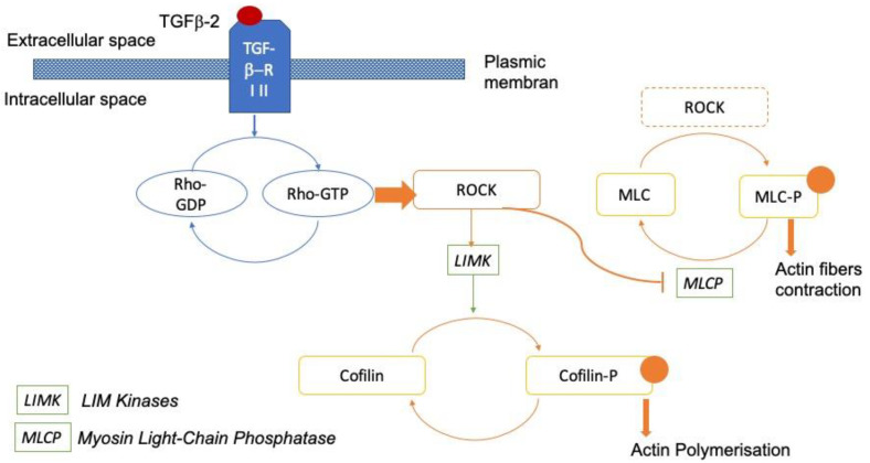
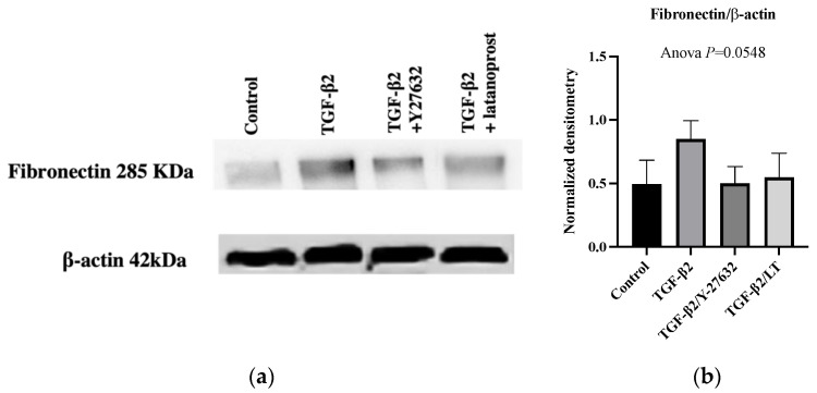
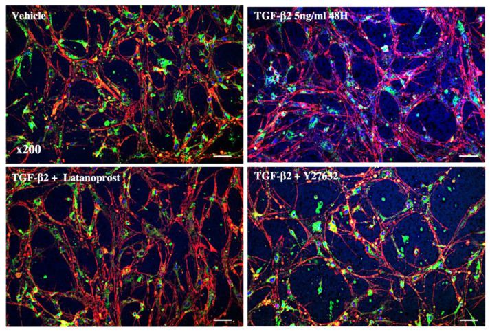
The trabecular meshwork (TM) is the main site of drainage of the aqueous humor, and its dysfunction leads to intraocular pressure elevation, which is one of the main risk factors of glaucoma. We aimed to compare the effects on cytoskeleton organization and extracellular matrix (ECM) of latanoprost (LT) and a Rho-kinase inhibitor (ROCKi) on a transforming growth factor beta2 (TGF-β2)-induced glaucoma-like model developed from primary culture of human TM cells (pHTMC). The TGF-β2 stimulated pHTMC were grown and incubated with LT or a ROCKi (Y-27632) for 24 h. The expression of alpha-smooth muscle actin (αSMA) and fibronectin (FN), and phosphorylation of the myosin light chain (MLC-P) and Cofilin (Cofilin-P) were evaluated using immunofluorescence and Western blot. The architectural modifications were studied in a MatrigelTM 3D culture. TGF-β2 increased the expression of αSMA and FN in pHTMC and modified the cytoskeleton with cross-linked actin network formation. LT did not alter the expression of αSMA but decreased FN deposition. The ROCKi decreased TGF-β2-induced αSMA and FN expression, as well as MLC-P and Cofilin-P, and stimulated the cells to recover a basal cytoskeletal arrangement. In the preliminary 3D study, pHTMC organized in a mesh conformation showed the widening of the TM under the effect of Y-27632. By simultaneously modifying the organization of the cytoskeleton and the ECM, with fibronectin deposition and overexpression, TGF-β2 reproduced the trabecular degeneration described in glaucoma. The ROCKi was able to reverse the TGF-β2-induced cytoskeletal and ECM rearrangements. LT loosened the extracellular matrix but had no action on the stress fibers.
Keywords: 3D culture; Matrigel; cytoskeleton; glaucoma; intraocular pressure; outflow; prostaglandin analog; rho-kinase inhibitor; trabecular meshwork.
Conflict of interest statement
Christophe Baudouin: Financial support and consultant (Alcon; Allergan; Santen; Laboratoires Théa). The other authors declare no conflict of interest.
Figures


Similar articles
-
Inhibition of TGF-β2-Induced Trabecular Meshwork Fibrosis by Pirfenidone.Transl Vis Sci Technol. 2023 Nov 1;12(11):21. doi: 10.1167/tvst.12.11.21. Transl Vis Sci Technol. 2023. PMID: 37975842 Free PMC article.
-
Mechanistic basis of Rho GTPase-induced extracellular matrix synthesis in trabecular meshwork cells.Am J Physiol Cell Physiol. 2010 Mar;298(3):C749-63. doi: 10.1152/ajpcell.00317.2009. Epub 2009 Nov 25. Am J Physiol Cell Physiol. 2010. PMID: 19940066 Free PMC article.
-
Effects of TGF-beta2, BMP-4, and gremlin in the trabecular meshwork: implications for glaucoma.Invest Ophthalmol Vis Sci. 2007 Mar;48(3):1191-200. doi: 10.1167/iovs.06-0296. Invest Ophthalmol Vis Sci. 2007. PMID: 17325163
-
The role of TGF-β2 and bone morphogenetic proteins in the trabecular meshwork and glaucoma.J Ocul Pharmacol Ther. 2014 Mar-Apr;30(2-3):154-62. doi: 10.1089/jop.2013.0220. Epub 2014 Feb 11. J Ocul Pharmacol Ther. 2014. PMID: 24517218 Free PMC article. Review.
-
The role of TGF-β in the pathogenesis of primary open-angle glaucoma.Cell Tissue Res. 2012 Jan;347(1):279-90. doi: 10.1007/s00441-011-1274-7. Epub 2011 Nov 19. Cell Tissue Res. 2012. PMID: 22101332 Review.
Cited by
-
Application of Single Cell Type-Derived Spheroids Generated by Using a Hanging Drop Culture Technique in Various In Vitro Disease Models: A Narrow Review.Cells. 2024 Sep 14;13(18):1549. doi: 10.3390/cells13181549. Cells. 2024. PMID: 39329734 Free PMC article. Review.
-
Glaucoma: Novel antifibrotic therapeutics for the trabecular meshwork.Eur J Pharmacol. 2023 Sep 5;954:175882. doi: 10.1016/j.ejphar.2023.175882. Epub 2023 Jun 28. Eur J Pharmacol. 2023. PMID: 37391006 Free PMC article. Review.
-
Inhibition of TGF-β2-Induced Trabecular Meshwork Fibrosis by Pirfenidone.Transl Vis Sci Technol. 2023 Nov 1;12(11):21. doi: 10.1167/tvst.12.11.21. Transl Vis Sci Technol. 2023. PMID: 37975842 Free PMC article.
-
Evaluation of Rho kinase inhibitor effects on neuroprotection and neuroinflammation in an ex-vivo retinal explant model.Acta Neuropathol Commun. 2024 Sep 14;12(1):150. doi: 10.1186/s40478-024-01859-z. Acta Neuropathol Commun. 2024. PMID: 39300576 Free PMC article.
-
Effects of a ROCK Inhibitor on Retinal Ganglion Cells In Vivo and In Vitro.J Clin Med. 2025 Jul 29;14(15):5344. doi: 10.3390/jcm14155344. J Clin Med. 2025. PMID: 40806966 Free PMC article.
References
-
- Kass M.A., Heuer D.K., Higginbotham E.J., Johnson C.A., Keltner J.L., Miller J.P., Parrish R.K., Wilson M.R., Gordon M.O. The Ocular Hypertension Treatment Study: A Randomized Trial Determines That Topical Ocular Hypotensive Medication Delays or Prevents the Onset of Primary Open-Angle Glaucoma. Arch. Ophthalmol. 2002;120:701–713; discussion 829–830. doi: 10.1001/archopht.120.6.701. - DOI - PubMed
LinkOut - more resources
Full Text Sources
Miscellaneous
