

Expanding the Role of Ultrasound for the Characterization of Renal Masses
- PMID: 35207384
- PMCID: PMC8876198
- DOI: 10.3390/jcm11041112
Expanding the Role of Ultrasound for the Characterization of Renal Masses
Abstract
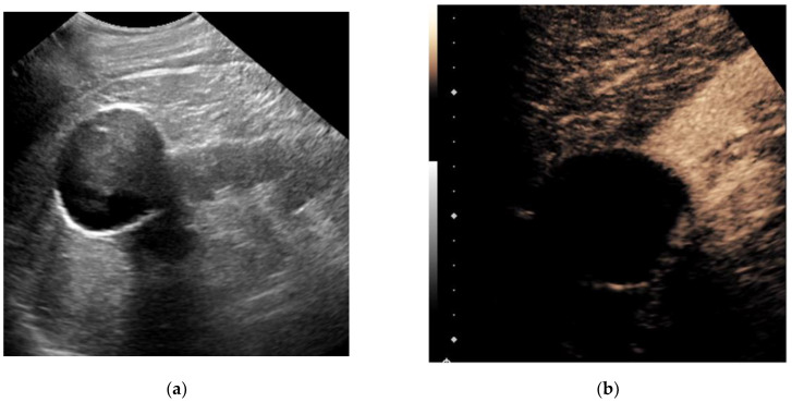
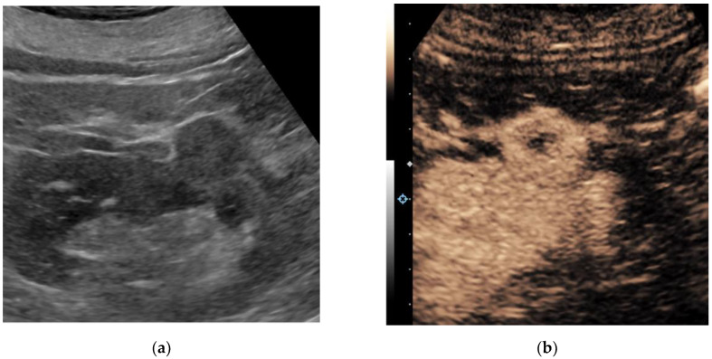
The incidental detection of renal masses has been steadily rising. As a significant proportion of renal masses that are surgically treated are benign or indolent in nature, there is a clear need for better presurgical characterization of renal masses to minimize unnecessary harm. Ultrasound is a widely available and relatively inexpensive real-time imaging technique, and novel ultrasound-based applications can potentially aid in the non-invasive characterization of renal masses. Evidence acquisition: We performed a narrative review on novel ultrasound-based techniques that can aid in the non-invasive characterization of renal masses. Evidence synthesis: Contrast-enhanced ultrasound (CEUS) adds significant diagnostic value, particularly for cystic renal masses, by improving the characterization of fine septations and small nodules, with a sensitivity and specificity comparable to magnetic resonance imaging (MRI). Additionally, the performance of CEUS for the classification of benign versus malignant renal masses is comparable to that of computed tomography (CT) and MRI, although the imaging features of different tumor subtypes overlap significantly. Ultrasound molecular imaging with targeted contrast agents is being investigated in preclinical research as an addition to CEUS. Elastography for the assessment of tissue stiffness and micro-Doppler imaging for the improved detection of intratumoral blood flow without the need for contrast are both being investigated for the characterization of renal masses, though few studies have been conducted and validation is lacking. Conclusions: Several novel ultrasound-based techniques have been investigated for the non-invasive characterization of renal masses. CEUS has several advantages over traditional grayscale ultrasound, including the improved characterization of cystic renal masses and the potential to differentiate benign from malignant renal masses to some extent. Ultrasound molecular imaging offers promise for serial disease monitoring and the longitudinal assessment of treatment response, though this remains in the preclinical stages of development. While elastography and emerging micro-Doppler techniques have shown some encouraging applications, they are currently not ready for widespread clinical use.
Keywords: contrast-enhanced ultrasound; micro-Doppler; molecular ultrasound; renal cell carcinoma; renal mass; renal tumor; ultrasonography; ultrasound.
Conflict of interest statement
Eduard Roussel has received unconditional research grants from Ipsen and Pfizer. Alexandre Ingels has received honoraria from Intuitive Surgical, Ipsen and Bristol Myers Squibb. Michele Marchioni has received honoraria from Astellas and Ipsen. Benoit Beuselinck received an unrestricted research grant from Bristol Myers Squibb and honoraria from Merck, Pfizer, Bristol Myers Squibb, Ipsen and AstraZeneca, and is a senior clinical investigator of “Fonds voor wetenschappelijk onderzoek Vlaanderen” (Belgium). David Fetzer received research support from GE Healthcare, Philips Healthcare and Siemens Healthineers, and is a consultant to Philips Healthcare. Maarten Albersen received unrestricted research grants from Ipsen and Pfizer and is a clinical investigator of “Stichting tegen kanker” (Belgium). All other authors have nothing to disclose.
Figures
Similar articles
-
Renal Complex Cystic Masses: Usefulness of Contrast-Enhanced Ultrasound (CEUS) in Their Assessment and Its Agreement with Computed Tomography.Curr Urol Rep. 2016 Dec;17(12):89. doi: 10.1007/s11934-016-0646-7. Curr Urol Rep. 2016. PMID: 27787749
-
Contrast-enhanced ultrasound (CEUS) of cystic and solid renal lesions: a review.Abdom Imaging. 2015 Aug;40(6):1982-96. doi: 10.1007/s00261-015-0348-5. Abdom Imaging. 2015. PMID: 25588715 Review.
-
Comparison of the Diagnostic Performance of Contrast-enhanced Ultrasound with That of Contrast-enhanced Computed Tomography and Contrast-enhanced Magnetic Resonance Imaging in the Evaluation of Renal Masses: A Systematic Review and Meta-analysis.Eur Urol Oncol. 2020 Aug;3(4):464-473. doi: 10.1016/j.euo.2019.08.013. Epub 2019 Sep 27. Eur Urol Oncol. 2020. PMID: 31570270
-
Multislice computed tomography versus contrast-enhanced ultrasound in evaluation of complex cystic renal masses using the Bosniak classification system.Clin Hemorheol Microcirc. 2008;39(1-4):171-8. Clin Hemorheol Microcirc. 2008. PMID: 18503122
-
The added value of contrast-enhanced ultrasound in evaluation of indeterminate small solid renal masses and risk stratification of cystic renal lesions.Eur Radiol. 2021 Nov;31(11):8468-8477. doi: 10.1007/s00330-021-07964-0. Epub 2021 Apr 29. Eur Radiol. 2021. PMID: 33912992
Cited by
-
Renal cell carcinoma of different pathological types in bilateral native kidneys of a kidney transplant recipient: A case report and literature review.Front Oncol. 2023 Jan 16;12:1112343. doi: 10.3389/fonc.2022.1112343. eCollection 2022. Front Oncol. 2023. PMID: 36727063 Free PMC article.
-
Artificial Intelligence in Renal Cell Carcinoma Histopathology: Current Applications and Future Perspectives.Diagnostics (Basel). 2023 Jul 6;13(13):2294. doi: 10.3390/diagnostics13132294. Diagnostics (Basel). 2023. PMID: 37443687 Free PMC article. Review.
-
A comparison of effectiveness of the contrast enhanced computed tomography with magnetic resonance imaging in the differential diagnosis of clear cell renal carcinoma.Radiol Oncol. 2025 Jun 16;59(2):193-202. doi: 10.2478/raon-2025-0033. eCollection 2025 Jun 1. Radiol Oncol. 2025. PMID: 40544505 Free PMC article.
-
Characteristics of high frame frequency contrast-enhanced ultrasound in renal tumors.BMC Med Imaging. 2024 Mar 25;24(1):71. doi: 10.1186/s12880-024-01245-0. BMC Med Imaging. 2024. PMID: 38528467 Free PMC article.
-
Correlation Between BMI and Kidney Tumor Lateralization: Insights into Survival and Risk Factors.Cancers (Basel). 2024 Dec 12;16(24):4139. doi: 10.3390/cancers16244139. Cancers (Basel). 2024. PMID: 39766039 Free PMC article.
References
-
- Johnson D.C., Vukina J., Smith A.B., Meyer A.-M., Wheeler S.B., Kuo T.-M., Tan H.-J., Woods M.E., Raynor M.C., Wallen E.M., et al. Preoperatively Misclassified, Surgically Removed Benign Renal Masses: A Systematic Review of Surgical Series and United States Population Level Burden Estimate. J. Urol. 2015;193:30–35. doi: 10.1016/j.juro.2014.07.102. - DOI - PubMed
-
- Marconi L., Dabestani S., Lam T.B., Hofmann F., Stewart F., Norrie J., Bex A., Bensalah K., Canfield S.E., Hora M., et al. Systematic Review and Meta-analysis of Diagnostic Accuracy of Percutaneous Renal Tumour Biopsy. [(accessed on 23 December 2021)];Eur. Urol. 2016 69:660–673. doi: 10.1016/j.eururo.2015.07.072. Available online: http://www.sciencedirect.com/science/article/pii/S0302283815007411. - DOI - PubMed
-
- Patel H.D., Johnson M.H., Pierorazio P.M., Sozio S., Sharma R., Iyoha E., Bass E., Allaf M.E. Diagnostic Accuracy and Risks of Biopsy in the Diagnosis of a Renal Mass Suspicious for Localized Renal Cell Carcinoma: Systematic Review of the Literature. J. Urol. 2016;195:1340–1347. doi: 10.1016/j.juro.2015.11.029. - DOI - PMC - PubMed
-
- Nientiedt M., Bertolo R., Campi R., Capitanio U., Erdem S., Kara Ö., Klatte T., Larcher A., Mir M.C., Ouzaid I., et al. Chronic Kidney Disease after Partial Nephrectomy in Patients with Preoperative Inconspicuous Renal Function—Curiosity or Relevant Issue? Clin. Genitourin. Cancer. 2020;18:e754–e761. doi: 10.1016/j.clgc.2020.05.007. - DOI - PubMed
Publication types
LinkOut - more resources
Full Text Sources
Research Materials
