

A Novel Anti-B7-H3 × Anti-CD3 Bispecific Antibody with Potent Antitumor Activity
- PMID: 35207448
- PMCID: PMC8879513
- DOI: 10.3390/life12020157
A Novel Anti-B7-H3 × Anti-CD3 Bispecific Antibody with Potent Antitumor Activity
Abstract
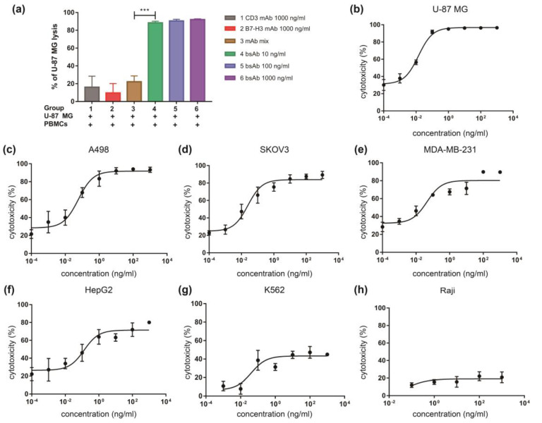
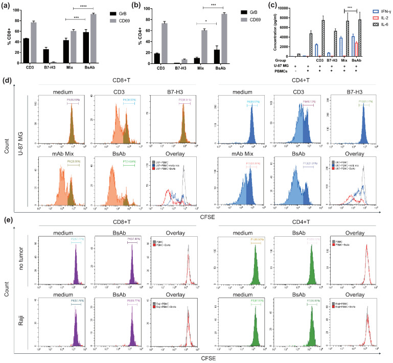
B7-H3 plays an important role in tumor apoptosis, proliferation, adhesion, angiogenesis, invasion, migration, and evasion of immune surveillance. It is overexpressed in various human solid tumor tissues. In patients, B7-H3 overexpression correlates with advanced stages, poor clinical outcomes, and resistance to therapy. The roles of B7-H3 in tumor progression make it a potential candidate for targeted therapy. Here, we generated a mouse anti-human B7-H3 antibody and demonstrated its binding activity via Tongji University Suzhou Instituteprotein-based and cell-based assays. We then developed a novel format anti-B7-H3 × anti-CD3 bispecific antibody based on the antibody-binding fragment of the anti-B7-H3 antibody and single-chain variable fragment structure of anti-CD3 antibody (OKT3) and demonstrated that this bispecific antibody mediated potent cytotoxic activities against various B7-H3-positive tumor cell lines in vitro by improving T cell activation and proliferation. This bispecific antibody also demonstrated potent antitumor activity in humanized mice xenograft models. These results revealed that the novel anti-B7-H3 × anti-CD3 bispecific antibody has the potential to be employed in treatment of B7-H3-positive solid tumors.
Keywords: B7-H3; bispecific T cell engager; immunotherapy; solid tumor.
Conflict of interest statement
The authors declare no conflict of interest.
Figures


Similar articles
-
Bispecific anti-CD3 x anti-B7-H3 antibody mediates T cell cytotoxic ability to human melanoma in vitro and in vivo.Invest New Drugs. 2019 Oct;37(5):1036-1043. doi: 10.1007/s10637-018-00719-7. Epub 2019 Feb 1. Invest New Drugs. 2019. PMID: 30706335
-
A T-cell-engaging B7-H4/CD3-bispecific Fab-scFv Antibody Targets Human Breast Cancer.Clin Cancer Res. 2019 May 1;25(9):2925-2934. doi: 10.1158/1078-0432.CCR-17-3123. Epub 2019 Feb 8. Clin Cancer Res. 2019. PMID: 30737243
-
MEK Inhibitor Augments Antitumor Activity of B7-H3-Redirected Bispecific Antibody.Front Oncol. 2020 Aug 25;10:1527. doi: 10.3389/fonc.2020.01527. eCollection 2020. Front Oncol. 2020. PMID: 32984002 Free PMC article.
-
Targeting B7-H3-A Novel Strategy for the Design of Anticancer Agents for Extracranial Pediatric Solid Tumors Treatment.Molecules. 2023 Apr 11;28(8):3356. doi: 10.3390/molecules28083356. Molecules. 2023. PMID: 37110590 Free PMC article. Review.
-
B7-H3: An Attractive Target for Antibody-based Immunotherapy.Clin Cancer Res. 2021 Mar 1;27(5):1227-1235. doi: 10.1158/1078-0432.CCR-20-2584. Epub 2020 Oct 13. Clin Cancer Res. 2021. PMID: 33051306 Free PMC article. Review.
Cited by
-
B7-H3 in Pediatric Tumors: Far beyond Neuroblastoma.Cancers (Basel). 2023 Jun 21;15(13):3279. doi: 10.3390/cancers15133279. Cancers (Basel). 2023. PMID: 37444389 Free PMC article. Review.
-
B7 Family Members in Pancreatic Ductal Adenocarcinoma: Attractive Targets for Cancer Immunotherapy.Int J Mol Sci. 2022 Nov 30;23(23):15005. doi: 10.3390/ijms232315005. Int J Mol Sci. 2022. PMID: 36499340 Free PMC article. Review.
-
Potent Apoptosis Induction by a Novel Trispecific B7-H3xCD16xTIGIT 2+1 Common Light Chain Natural Killer Cell Engager.Molecules. 2024 Mar 4;29(5):1140. doi: 10.3390/molecules29051140. Molecules. 2024. PMID: 38474651 Free PMC article.
-
METTL3 inhibition induced by M2 macrophage-derived extracellular vesicles drives anti-PD-1 therapy resistance via M6A-CD70-mediated immune suppression in thyroid cancer.Cell Death Differ. 2023 Oct;30(10):2265-2279. doi: 10.1038/s41418-023-01217-x. Epub 2023 Aug 30. Cell Death Differ. 2023. PMID: 37648786 Free PMC article.
-
B7-H3 in acute myeloid leukemia: From prognostic biomarker to immunotherapeutic target.Chin Med J (Engl). 2024 Nov 5;137(21):2540-2551. doi: 10.1097/CM9.0000000000003099. Epub 2024 Apr 9. Chin Med J (Engl). 2024. PMID: 38595093 Free PMC article. Review.
References
Grants and funding
LinkOut - more resources
Full Text Sources
Research Materials
