. 2022 Jan 29;12(2):208.
doi: 10.3390/life12020208.

Bim Expression Promotes the Clearance of Mononuclear Phagocytes during Choroidal Neovascularization, Mitigating Scar Formation in Mice

Affiliations

Bim Expression Promotes the Clearance of Mononuclear Phagocytes during Choroidal Neovascularization, Mitigating Scar Formation in Mice

Shoujian Wang et al. Life (Basel). .

Abstract

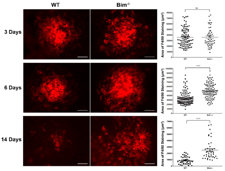
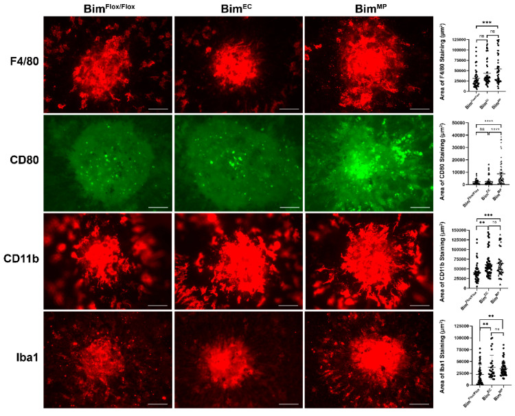
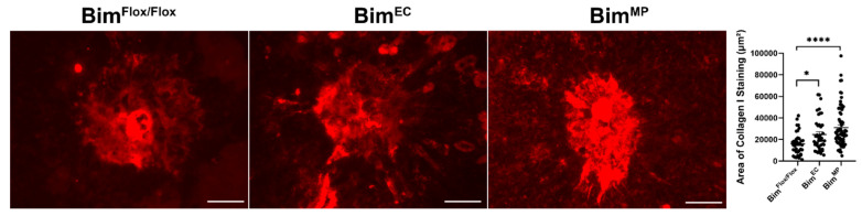
Inflammation is increasingly recognized as an important modulator in the pathogenesis of neovascular age-related macular degeneration (nAMD). Although significant progress has been made in delineating the pathways that contribute to the recruitment of inflammatory cells and their contribution to nAMD, we know little about what drives the resolution of these inflammatory responses. Gaining a better understanding of how immune cells are cleared in the choroid will give a novel insight into how sustained inflammation could influence the pathogenesis of nAMD. The pro-apoptotic Bcl-2 family member Bim is a master regulator of immune cell homeostasis. In its absence, immune cell lifespan and numbers increase. Most therapeutic regimes that squelch inflammation do so by enhancing immune cell apoptosis through enhanced Bim expression and activity. To test the hypothesis that Bim expression tempers inflammation during the pathogenesis of nAMD, we used the mouse laser-induced choroidal neovascularization (CNV) model in which inflammation acts as a facilitator of CNV. Here, we showed minimal to no change in the recruitment of F4/80-, CD80-, CD11b-, and Iba1-positive myeloid-derived mononuclear phagocytes to the site of laser photocoagulation in the absence of Bim expression. However, the resolution of these cells from the choroid of Bim-deficient (Bim -/-) mice was significantly diminished following laser photocoagulation. With time, we noted increased scar formation, demonstrated by collagen I staining, in Bim -/- mice with no change in the resolution of neovascularization compared to wild-type littermates. We also noted that mice lacking Bim expression in mononuclear phagocytes (BimFlox/Flox; Lyz2-Cre (BimMP) mice) had delayed resolution of F4/80-, CD80-, CD11b-, and Iba1-positive cells, while those lacking Bim expression in endothelial cells (BimFlox/Flox; Cad5-Cre (BimEC) mice) had delayed resolution of only CD11b- and Iba1-positive cells. Both BimMP and BimEC mice demonstrated increased scar formation, albeit to differing degrees. Thus, our studies show that resolving inflammation plays an important role in moderating scar formation in nAMD, and it is impacted by Bim expression in both the endothelium and mononuclear phagocyte lineages.

Keywords: Bcl-2 family; age-related macular degeneration; angiogenesis; choroid; inflammation; macrophages; vascular dysfunction.


Conflict of interest statement

The authors state that the work presented here was not influenced by any commercial or financial relationships that could be considered as a potential conflict of interest.

Figures

Figure 1
Figure 1
Decreased resolution of F4/80 cells in the absence of Bim expression. Choroidal neovascularization was induced in 3-month-old wild-type (WT) and Bim -/- mice by laser-photocoagulation-induced rupture of Bruch’s membrane. At 3, 6, and 14 days following laser photocoagulation, the eyes were sectioned at the equator and the anterior half with the vitreous and retina discarded. The remaining RPE/choroid complex was stained with anti-F4/80, and the positive staining area quantified (ns; not significant; **** p < 0.0001). Each point represents one laser spot. Scale bar = 100 µm. These experiments were repeated with at least 8 mice of each genotype.
Figure 2
Figure 2
Increased CD11b-positive cells at 6 days following laser photocoagulation in Bim -/- mice. RPE/choroid complex was harvested from 3-month-old wild-type and Bim -/- mice 3, 6, and 14 days following laser photocoagulation. The RPE/choroid complex was wholemount stained with anti-CD11b and the positive staining area quantified (**** p < 0.0001). Each point represents one laser spot. Scale bar = 100 µm. These experiments were repeated with at least 8 mice of each genotype.
Figure 3
Figure 3
Increased number of CD11b-positive cells in Bim -/- naïve choroid. RPE/choroid complex was harvested from naïve 8-week-old wild-type (WT) and Bim -/- mice. The complex was wholemount stained with anti-CD11b and the positive staining area quantified. Each point represents one RPE/choroid complex. These experiments were repeated with at least 6 mice of each genotype. Scale bar = 50 µm. (**** p < 0.0001).
Figure 4
Figure 4
Decreased resolution of CD80-positive cells in RPE/choroid from Bim -/- mice. Three-month-old wild-type and Bim -/- mice underwent laser photocoagulation. The choroid RPE/choroid complex was harvested after 3, 6, and 14 days, wholemount stained with anti-CD80, and the positive staining area was quantified. Each point represents one laser spot. These experiments were repeated with at least 8 mice of each genotype. Scale bar = 100 µm. (** p < 0.01, **** p < 0.0001).
Figure 5
Figure 5
Global lack of Bim expression increased Iba1-positive cells. Wild-type and Bim -/- mice (3-month-old) underwent laser photocoagulation, and the RPE/choroid was harvested 3, 6, and 14 days later. The RPE/choroid was wholemount stained with anti-Iba1 and the positive staining area was quantified. Each point represents one laser spot. These experiments were repeated with at least 8 mice from each genotype. Scale bar = 100 µm. (* p < 0.05, *** p < 0.001, **** p < 0.0001).
Figure 6
Figure 6
Mice lacking Bim expression have similar recruitment but prolonged resolution of MPs compared to WT counterparts. To further illustrate MP recruitment and clearance with time, the data in Figure 1, Figure 2, Figure 4 and Figure 5 were compiled into line graphs. ** p < 0.01, *** p < 0.001, **** p < 0.0001.
Figure 7
Figure 7
Mice conditionally lacking Bim expression in MPs have decreased resolution of F4/80-, CD80-, CD11b-, and Iba1-positive cells. To assess the role Bim expression plays in moderating the immune response during CNV, 3-month-old BimFlox/Flox, BimEC, and BimMP mice underwent laser photocoagulation. Six days later, the RPE/choroid was wholemount stained with anti-F4/80, anti-CD80, anti-CD11b, or anti-Iba1, and the area of positive staining was quantified (on the right). Each point represents one laser spot. Experiments were performed with at least 8 mice for each condition. Scale bar = 100 µm. (ns; not significant; **** p < 0.0001, *** p < 0.001, ** p < 0.01).
Figure 8
Figure 8
Increased collagen I in the absence of Bim. To assess the amount of scarring, the RPE/choroid from wild-type and Bim -/- mice was wholemount stained with anti-collagen I, 2 and 5 weeks following laser photocoagulation. The area of collagen staining was quantified (on the right). Each point represents one laser spot. At least 8 mice for each condition were used. Scale bar = 100 µm. (ns; not significant; ** p < 0.01).
Figure 9
Figure 9
Similar level of CNV was noted 5 weeks following laser photocoagulation in and wild type and Bim -/- mice. Wild-type and Bim -/- mice (3-month-old) underwent laser photocoagulation. Five weeks later, the RPE/choroid was harvested, wholemount stained with anti-ICAM-2, and the area of staining quantified. Each point represents one laser spot. At least 8 mice for each condition were used. Scale bar = 100 µm. (ns; not significant).
Figure 10
Figure 10
Increased scar accumulation in mice conditionally lacking Bim. Three-month-old BimFlox/Flox, BimEC, and BimMP mice were subjected to laser photocoagulation. Five weeks following laser photocoagulation, the RPE/choroid was harvested, stained with anti-collagen I, and the area of staining was quantified. Each point represents one laser spot. For each condition at least 8 mice were used. Scale bar = 100 µm. (* p < 0.05, **** p < 0.0001).

Similar articles

Cited by

References

    1. Jager R.D., Mieler W.F., Miller J.W. Age-related macular degeneration. N. Engl. J. Med. 2008;358:2606–2617. doi: 10.1056/NEJMra0801537. - DOI - PubMed
    1. Pemp B., Schmetterer L. Ocular blood flow in diabetes and age-related macular degeneration. Can. J. Ophthalmol. 2008;43:295–301. doi: 10.3129/i08-049. - DOI - PubMed
    1. Farnoodian M., Wang S., Dietz J., Nickells R.W., Sorenson C.M., Sheibani N. Negative regulators of angiogenesis: Important targets for treatment of exudative amd. Clin. Sci. 2017;131:1763–1780. doi: 10.1042/CS20170066. - DOI - PMC - PubMed
    1. Carmeliet P., Ferreira V., Breier G., Pollefeyt S., Kieckens L., Gertsenstein M., Fahrig M., Vandenhoeck A., Harpal K., Eberhardt C., et al. Abnormal blood vessel development and lethality in embryos lacking a single vegf allele. Nature. 1996;380:435–439. doi: 10.1038/380435a0. - DOI - PubMed
    1. Ferrara N., Carver-Moore K., Chen H., Dowd M., Lu L., O’Shea K.S., Powell-Braxton L., Hillan K.J., Moore M.W. Heterozygous embryonic lethality induced by targeted inactivation of the vegf gene. Nature. 1996;380:439–442. doi: 10.1038/380439a0. - DOI - PubMed

LinkOut - more resources