Review
. 2022 Jan 31;12(2):187.
doi: 10.3390/jpm12020187.

Diagnostic Challenges in Rare Causes of Arrhythmogenic Cardiomyopathy-The Role of Cardiac MRI

Affiliations
Review

Diagnostic Challenges in Rare Causes of Arrhythmogenic Cardiomyopathy-The Role of Cardiac MRI

Simona Manole et al. J Pers Med. .

Abstract

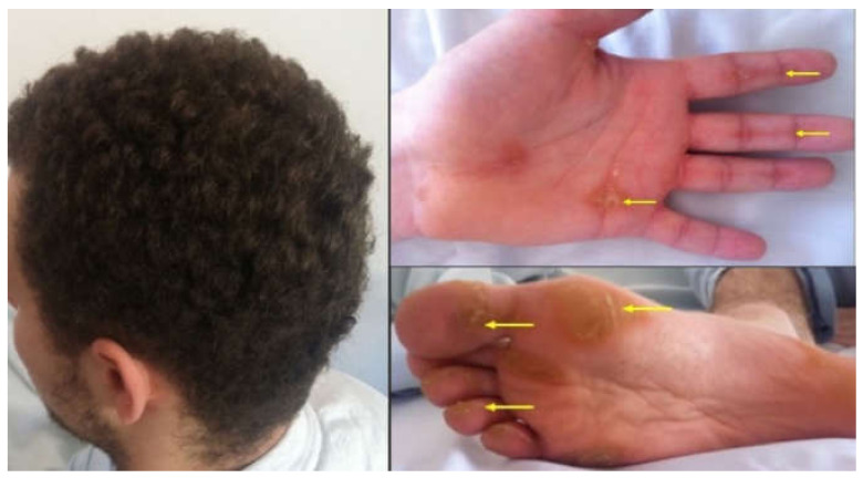
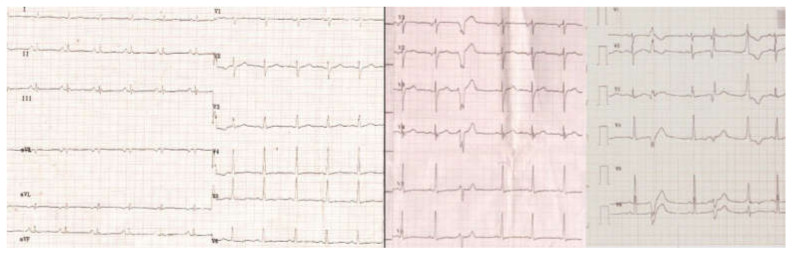
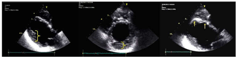
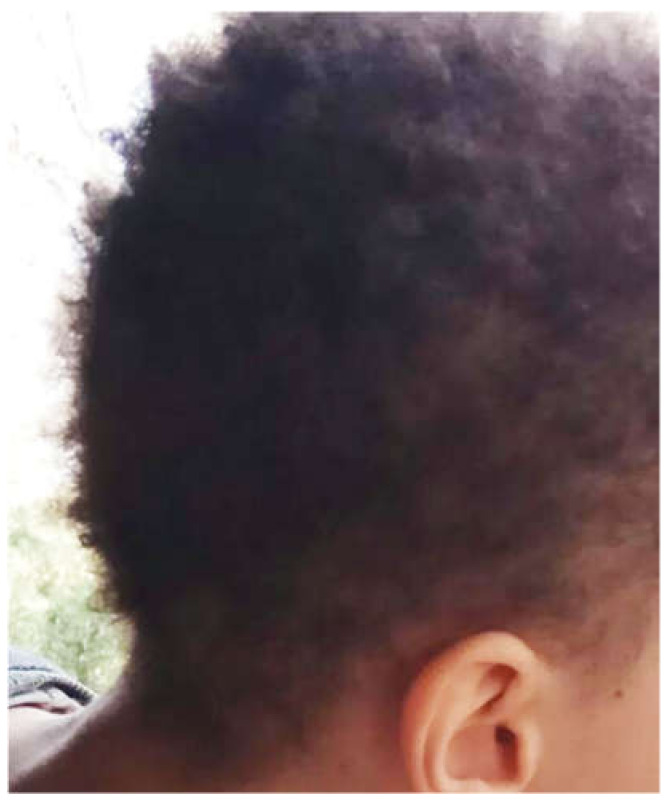
Arrhythmogenic right ventricular dysplasia (ARVD) is a rare genetic condition of the myocardium, with a significantly high risk of sudden death. Recent genetic research and improved understanding of the pathophysiology tend to change the ARVD definition towards a larger spectrum of myocardial involvement, which includes, in various proportions, both the right (RV) and left ventricle (LV), currently referred to as ACM (arrhythmogenic cardiomyopathy). Its pathological substrate is defined by the replacement of the ventricular myocardium with fibrous adipose tissue that further leads to inadequate electrical impulses and translates into varies degrees of malignant ventricular arrythmias and dyskinetic myocardium movements. Particularly, the cardio-cutaneous syndromes of Carvajal/Naxos represent rare causes of ACM that might be suspected from early childhood. The diagnostic is sometimes challenging, even with well-established rTFC or Padua criteria, especially for pediatric patients or ACM with LV involvement. Cardiac MRI gain more and more importance in ACM diagnostic especially in non-classical forms. Furthermore, MRI is useful in highlighting myocardial fibrosis, fatty replacement or wall movement with high accuracy, thus guiding not only the depiction, but also the patient's stratification and management.

Keywords: ACM; ACM-LV; Padua; arrhythmogenic cardiomyopathy; cardio-cutaneous syndrome.


Conflict of interest statement

The authors declare no conflict of interest.

Figures

Figure 1
Figure 1
The patient has woolly hair, characterized by the fact that the hair is curly and dull. Striated fibrous palmar hyperkeratosis—hyperkeratotic lesions are located on the fingers and flexion areas (arrows). Focal plantar hyperkeratosis—hyperkeratotic lesions are located in the pressure zones (arrows).
Figure 2
Figure 2
The ECG shows hypovoltage, nonspecific intraventricular conduction disturbances, negative T-waves in V5-V6 and polymorphic ventricular extrasystoles.
Figure 3
Figure 3
Cardiac ultrasound. Left image: long-axis parasternal section shows dilated LV, with hyperechoic thin SIV, and LV noncompaction areas at the posterolateral wall (brace); middle image: short axis parasternal section displays noncompact LV area (brace) > 2 × compact LV area; right image: parasternal section long axis shows aneurysms located at the SIV level (arrows).
Figure 4
Figure 4
(A) Axial Cine-GRE sequence: focal dyskinesia of the RVOT; (B) Cine-GRE two-chamber LV sequence: focal dyskinesia of the anterior wall of the LV; (C,D) four-chamber Cine-GRE sequence: focal dyskinesia of the RV apex (arrow), dyskinesia of the lateral wall of the LV (square brackets), akinetic IVS areas (black arrows); (E) PSIR sequence—delayed postcontrast axial view: concentric midmyocardial LGE within the LV and diffuse, inhomogeneous LGE of the lateral wall of RV; (F) PSIR sequence—delayed postcontrast short axis view: midmyocardial LGE of the LV base.
Figure 5
Figure 5
Woolly hair in a 10-y old patient.
Figure 6
Figure 6
(A) Short axis Cine-GRE sequence: basal dilatation of the LV, normal size RV; (B) short axis delayed postcontrast PSIR sequence: concentric midmyocardial LGE of LV; (C) LV two chambers delayed postcontrast PSIR sequence: non-uniform midmyocardial LGE of the anterior wall of the LV.
Figure 7
Figure 7
(A) Axial Cine-GRE sequence: focal dyskinesia of the RVOT; (B) four-chamber Cine-GRE sequence, systole: focal dyskinesias of the lateral wall of the RV, above the insertion of the moderating band; (C) PSIR delayed postcontrast sequence short-axis: diffuse LGE of the lateral wall of the RV, focal LGE of the inferior wall (arrow); (D) Axial delayed postcontrast PSIR sequence: LGE of the lateral wall of the RV.

Similar articles

Cited by

References

    1. Pilichou K., Thiene G., Bauce B. Arrhythmogenic cardiomyopathy. Orphanet J. Rare Dis. 2016;11:33. doi: 10.1186/s13023-016-0407-1. - DOI - PMC - PubMed
    1. Basso C., Pilichou K., Bauce B., Corrado D., Thiene G. Diagnostic Criteria, Genetics, and Molecular Basis of Arrhythmogenic Cardiomyopathy. Heart Fail Clin. 2018;14:201–213. doi: 10.1016/j.hfc.2018.01.002. - DOI - PubMed
    1. Lazzarini E., Jongbloed J.D., Pilichou K., Thiene G., Basso C., Bikker H., Charbon B., Swertz M., van Tintelen J.P., van der Zwaag P.A. The ARVD/C Genetic Variants Database: 2014 Update. Hum. Mutat. 2015;36:403–410. doi: 10.1002/humu.22765. - DOI - PubMed
    1. Bennett R.G., Haqqani H.M., Berruezo A., Della Bella P., Marchlinski F.E., Hsu C.-J., Kumar S. Arrhythmogenic Cardiomyopathy in 2018–2019: ARVC/ALVC or Both? Heart Lung Circ. 2019;28:164–177. doi: 10.1016/j.hlc.2018.10.013. - DOI - PubMed
    1. Protonotarios A., Elliott P.M. Arrhythmogenic Cardiomyopathy: A Disease or Merely a Phenotype? Eur. Cardiol. 2020;15:1–5. doi: 10.15420/ecr.2019.05. - DOI - PMC - PubMed

LinkOut - more resources