

Mechanism Assay of Honeysuckle for Heat-Clearing Based on Metabolites and Metabolomics
- PMID: 35208196
- PMCID: PMC8874459
- DOI: 10.3390/metabo12020121
Mechanism Assay of Honeysuckle for Heat-Clearing Based on Metabolites and Metabolomics
Abstract
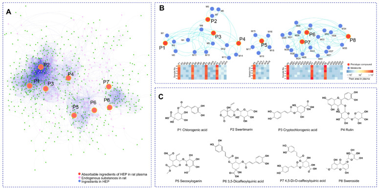
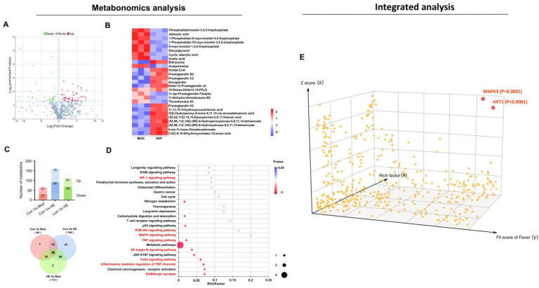
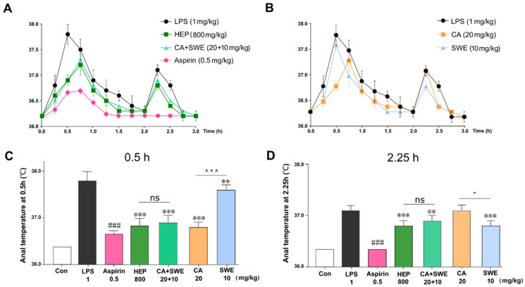
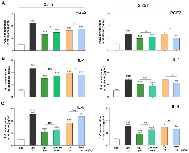
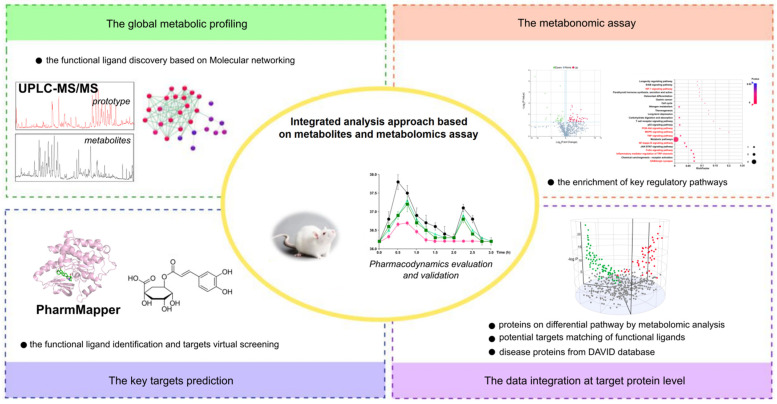
Nonsteroidal anti-inflammatory drugs (NSAIDs), such as cyclooxygenase (Cox)-1/2 inhibitor, have emerged as potent antipyretics and analgesics. However, few herbs with Cox-1/2 inhibitory activity are commonly used for heat-clearing in China. Although these are known to have antipyretic activity, there is a lack of molecular data supporting their activity. Using the traditional Chinese medicine herb honeysuckle (Hon) as an example, we explored key antipyretic active compounds and their mechanisms of action by assessing their metabolites and metabolomics. Mitogen-activated protein kinase (MAPK) 3 and protein kinase B (AKT) 1 were suggested as key targets regulated primarily by chlorogenic acid (CA) and swertiamarin (SWE). CA and SWE synergistically inhibited the production of interleukin (IL)-1 and IL-6, alleviated generation of prostaglandin E2, and played an antipyretic role equivalent to honeysuckle extract at the same dose contents within 3 h. Collectively, these findings indicated that lipopolysaccharide-induced fever can be countered by CA with SWE synergistically, allowing the substitution of a crude extract of complex composition with active compounds. Our findings demonstrated that, unlike the traditional NSAIDs, the Hon extract showed a remote and indirect mechanism for alleviating fever that depended on the phosphatidylinositol-3-kinase-AKT and MAPK pathways by regulating the principal mediator of inflammation.
Keywords: anti-inflammatory; antipyretic; chlorogenic acid; metabolites; metabolomics; swertiamarin.
Conflict of interest statement
The authors declare no conflict of interest.
Figures
Similar articles
-
Network pharmacology integrated with molecular docking reveals the common experiment-validated antipyretic mechanism of bitter-cold herbs.J Ethnopharmacol. 2021 Jun 28;274:114042. doi: 10.1016/j.jep.2021.114042. Epub 2021 Mar 26. J Ethnopharmacol. 2021. PMID: 33775806
-
Antipyretic, anti-inflammatory and analgesic activities of Periplaneta americana extract and underlying mechanisms.Biomed Pharmacother. 2020 Mar;123:109753. doi: 10.1016/j.biopha.2019.109753. Epub 2019 Dec 19. Biomed Pharmacother. 2020. PMID: 31865148
-
Antipyretic and anti-inflammatory activities of Thais luteostoma extracts and underlying mechanisms.Chin J Nat Med. 2015 Mar;13(3):192-8. doi: 10.1016/S1875-5364(15)30004-2. Chin J Nat Med. 2015. PMID: 25835363
-
Towards developing new strategies to reduce the adverse side-effects of nonsteroidal anti-inflammatory drugs.Clin Exp Nephrol. 2012 Feb;16(1):25-9. doi: 10.1007/s10157-011-0492-3. Epub 2011 Nov 1. Clin Exp Nephrol. 2012. PMID: 22038259 Review.
-
Dual acting anti-inflammatory drugs: a reappraisal.Pharmacol Res. 2001 Dec;44(6):437-50. doi: 10.1006/phrs.2001.0872. Pharmacol Res. 2001. PMID: 11735348 Review.
Cited by
-
A Comprehensive Quality Analysis of Different Colors of Medicinal and Edible Honeysuckle.Foods. 2023 Aug 20;12(16):3126. doi: 10.3390/foods12163126. Foods. 2023. PMID: 37628125 Free PMC article.
-
Chlorogenic acid: a review on its mechanisms of anti-inflammation, disease treatment, and related delivery systems.Front Pharmacol. 2023 Sep 13;14:1218015. doi: 10.3389/fphar.2023.1218015. eCollection 2023. Front Pharmacol. 2023. PMID: 37781708 Free PMC article. Review.
-
Network pharmacology integrated with experimental verification reveals the antipyretic characteristics and mechanism of Zi Xue powder.Pharm Biol. 2023 Dec;61(1):1512-1524. doi: 10.1080/13880209.2023.2287658. Epub 2023 Dec 9. Pharm Biol. 2023. PMID: 38069658 Free PMC article.
-
Mass spectrometry-based metabolomics for discovering active ingredients and exploring action mechanism of herbal medicine.Front Chem. 2023 Mar 31;11:1142287. doi: 10.3389/fchem.2023.1142287. eCollection 2023. Front Chem. 2023. PMID: 37065828 Free PMC article. Review.
-
Investigating the Therapeutic Mechanisms of Honeysuckle (China) in Sepsis Through Network Pharmacology and Experimental Validation.Infect Drug Resist. 2025 Jul 3;18:3257-3277. doi: 10.2147/IDR.S499975. eCollection 2025. Infect Drug Resist. 2025. PMID: 40630747 Free PMC article.
References
Grants and funding
LinkOut - more resources
Full Text Sources
