. 2022 Feb 4;12(2):144.
doi: 10.3390/metabo12020144.

Integrative Analysis of Metabolome and Transcriptome Reveals the Mechanism of Color Formation in Liriope spicata Fruit

Affiliations

Integrative Analysis of Metabolome and Transcriptome Reveals the Mechanism of Color Formation in Liriope spicata Fruit

Sichen Gan et al. Metabolites. .

Abstract

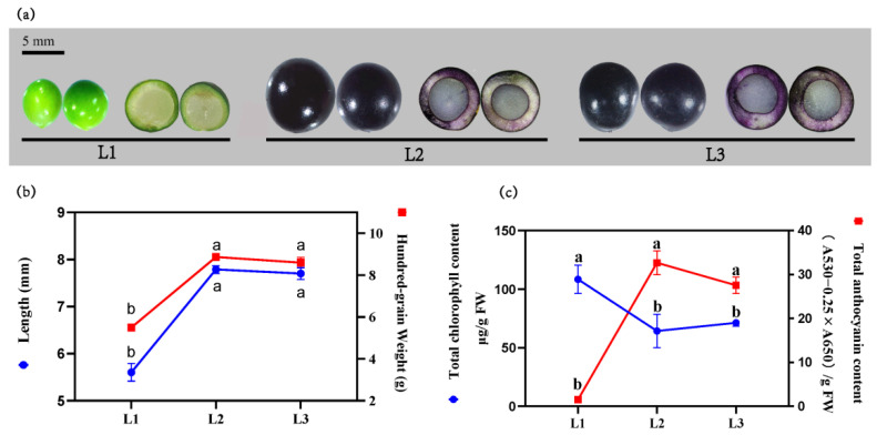
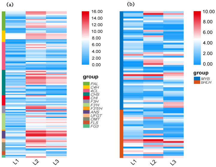
Liriope spicata is an important ornamental ground cover plant, with a fruit color that turns from green to black during the development and ripening stages. However, the material basis and regulatory mechanism of the color variation remains unclear. In this study, a total of 31 anthocyanins and 2 flavonols were identified from the skin of L. spicata fruit via integrative analysis on the metabolome and transcriptome of three developmental stages. The pigments of black/mature fruits are composed of five common anthocyanin compounds, of which Peonidin 3-O-rutinoside and Delphinidin 3-O-glucoside are the most differential metabolites for color conversion. Using dual-omics joint analysis, the mechanism of color formation was obtained as follows. The expression of structural genes including 4CL, F3H, F3'H, F3'5'H and UFGT were activated due to the upregulation of transcription factor genes MYB and bHLH. As a result, a large amount of precursor substances for the synthesis of flavonoids accumulated. After glycosylation, stable pigments were generated which promoted the accumulation of anthocyanins and the formation of black skin.

Keywords: Liriope spicata; anthocyanins; fruit skin; metabolome; transcriptome.


Conflict of interest statement

The authors declare no conflict of interest.

Figures

Figure 1
Figure 1
The dynamic variation of L. spicata fruits in morphology and pigment during three developmental stages. (a) The variation of fruit appearance, (b) length and hundred-grain weight, (c) TCC and TAC. Error bars represent ± SE of biological replicates (ten biological replicates × three stages, n = 30). Different lowercased letters in the same panel indicate statistical significance using Duncan’s test (p < 0.05).
Figure 2
Figure 2
Metabolite analysis of L. spicata fruits in different stages. (a) The content variation of anthocyanins and flavonol in L. spicata fruits. Error bars represent ± SE of biological replicates (three biological replicates × three stages, n = 9). (b) The heat map of DAMs during three stages. Intensity value bars are shown on the right side of the heat map. Colors indicate the normalized intensity of DAMs, of which the red color represents high content and blue corresponds to low content.
Figure 3
Figure 3
Heat map of DEGs. (a) The dynamic profile of differentially expressed structural genes and (b) regulation genes. Intensity value bars are shown on the right side of each heat map. Colors indicate the normalized intensity of DEGs. Red represents high expression and blue corresponds to low expression.
Figure 4
Figure 4
Phylogenetic analysis of (a) LsMYBs and (b) LsbHLHs TFs from L. spicata and compared with that of other species. Black nodes indicated LsMYBs and LsMYBs. Functions of all MYBs and bHLHs were listed on the right. Gene bank accession numbers of the used sequences were listed in Supplementary Table S3.
Figure 5
Figure 5
Relationship between module eigengenes (MEs, rows) and traits (columns). MEs were used to create the correlation and its p value between modules and traits (24 DAMs). The right panel described a color scale for module–DAMs correlations from –1 to 1.
Figure 6
Figure 6
Construction of regulatory networks of anthocyanin biosynthesis. Eight anthocyanins and thirty-six genes were applied to construct the network by means of Cytoscape (3.7.2 version) According to the type of anthocyanins to which genes related, 7 groups were created. The genes located in Group 1 to 7 corresponded to 8, 3, 6, 7, 1, 2, 5 types of anthocyanins, respectively.
Figure 7
Figure 7
RT–qPCR results of eleven randomly selected hub genes from darkorange modules, bisque4 modules and the anthocyanin–gene correlation network. Error bars represent ± SE of biological replicates (three biological replicates × three stages, n = 9).

References

    1. Tanaka Y., Sasaki N., Ohmiya A. Biosynthesis of plant pigments: Anthocyanins, betalains and carotenoids. Plant J. 2008;54:733–749. doi: 10.1111/j.1365-313X.2008.03447.x. - DOI - PubMed
    1. Hong H.T., Netzel M.E., O’Hare T.J. Anthocyanin composition and changes during kernel development in purple-pericarp supersweet sweetcorn. Food Chem. 2020;315:126284. doi: 10.1016/j.foodchem.2020.126284. - DOI - PubMed
    1. Lou Q., Liu Y., Qi Y., Jiao S., Tian F., Jiang L., Wang Y. Transcriptome sequencing and metabolite analysis reveals the role of delphinidin metabolism in flower colour in grape hyacinth. J. Exp. Bot. 2014;65:3157–3164. doi: 10.1093/jxb/eru168. - DOI - PMC - PubMed
    1. Tatsuzawa F., Tanikawa N., Nakayama M. Red-purple flower color and delphinidin-type pigments in the flowers of Pueraria lobata (Leguminosae) Phytochemistry. 2017;137:52–56. doi: 10.1016/j.phytochem.2017.02.004. - DOI - PubMed
    1. Yuhui Z.H.A.I., Jiaqi L., Xiang L.I., Xiaoning L.U.O., Long L.I., Qianqian S.H.I. Effects of cell sap pH on the flower color formation in Primula vulgaris. Acta Hortic. Sin. 2020;47:477–491.

LinkOut - more resources