

Atypical Sites of Lymphadenopathy after Anti-COVID-19 Vaccine: Ultrasound Features
- PMID: 35208521
- PMCID: PMC8878753
- DOI: 10.3390/medicina58020197
Atypical Sites of Lymphadenopathy after Anti-COVID-19 Vaccine: Ultrasound Features
Abstract
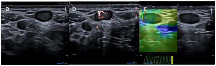
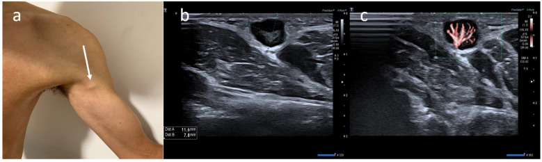
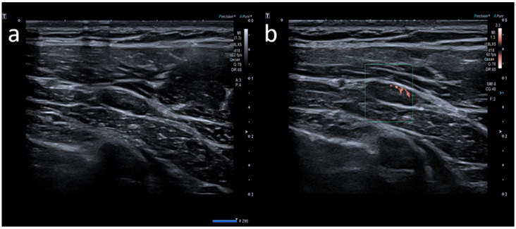
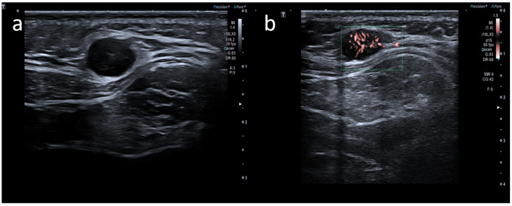
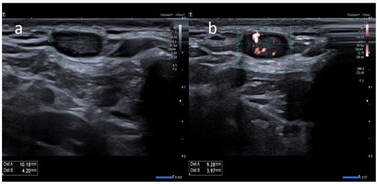
Background and Objectives: Several authors have reported cervical and axillary lymphadenopathies as known side effects following anti-COVID-19 vaccine administration. Few data are available about atypical locations of post-anti-COVID-19 vaccine lymphadenopathy. In this investigation, we evaluated the incidence and prevalence of postvaccine lymphadenopathy ultrasound (US) features in atypical sites. Materials and Methods: In this retrospective study, we retrospectively selected 64 patients on whom US was performed between January and October 2021 due to COVID-19 vaccine-related lymphadenopathy. We investigated lymph node anatomical sites, presence, number, size, shape, cortical profile, hilum outline, superb microvascular imaging (SMI), and elastosonography. Results: A total of 170 nodes were assessed. Atypical location was demonstrated in 5/64 patients (7.8%). In all these cases, atypical nodal involvement was associated with lymphadenopathy in a typical site (axillary, supraclavicular) ipsilateral to the vaccine injection site. Two patients presented lymphadenopathy in the infraclavicular station (3.1%), one in the pectoralis major muscle (1.6%), one in the left arm (1.6%), and one in the nuchal site (1.6%). All lymphadenopathies were oval-shaped, with a median size of 0.9 ± 0.2 cm. US features included a symmetric cortex with hilum evidence (4/6, 60%), vascular signal at SMI in both the hilar region and periphery of lymph node (5/6, 83.3%), and a US elastography pattern resembling that of adjacent tissues (5/6, 83.3%). The median age of patients with lymphadenopathies in an atypical location was 23 years. The main type of vaccine associated with lymph node appearance in atypical sites was Moderna's mRNA-1273 (60% of patients, 4/6 lymph nodes accounting for 66.7% among atypical locations). Conclusion: Post-COVID-19 vaccine administration lymphadenopathies in an atypical location represent an intense immune response to antigenic stimuli and they may show alarming US traits superimposed on malignant pathologies, which may complicate the patient's clinical and diagnostic pathway. Despite no distinctive US features between reactive post-COVID-19 vaccination and malignant lymph nodes being available, careful examination of atypical lymph node locations associated with accurate knowledge of patients' clinical background and delay of US exam to four to six weeks after vaccine injection should be considered.
Keywords: anti-COVID-19 vaccine; atypical sites; lymphadenopathy; ultrasound.
Conflict of interest statement
The authors declare no conflict of interest.
Figures
References
-
- WHO Coronavirus (COVID-19) Dashboard. [(accessed on 13 October 2021)]. Available online: https://covid19.who.int/
-
- Konopka K.E., Nguyen T., Jentzen J.M., Rayes O., Schmidt C.J., Wilson A.M., Farver C.F., Myers J.L. Diffuse alveolar damage (DAD) resulting from coronavirus disease 2019 Infection is Morphologically Indistinguishable from Other Causes of DAD. Histopathology. 2020;77:570–578. doi: 10.1111/his.14180. - DOI - PMC - PubMed
MeSH terms
Substances
LinkOut - more resources
Full Text Sources
Medical
