

Renal Cell Carcinoma or Oncocytoma? The Contribution of Diffusion-Weighted Magnetic Resonance Imaging to the Differential Diagnosis of Renal Masses
- PMID: 35208545
- PMCID: PMC8878185
- DOI: 10.3390/medicina58020221
Renal Cell Carcinoma or Oncocytoma? The Contribution of Diffusion-Weighted Magnetic Resonance Imaging to the Differential Diagnosis of Renal Masses
Abstract
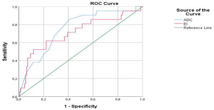
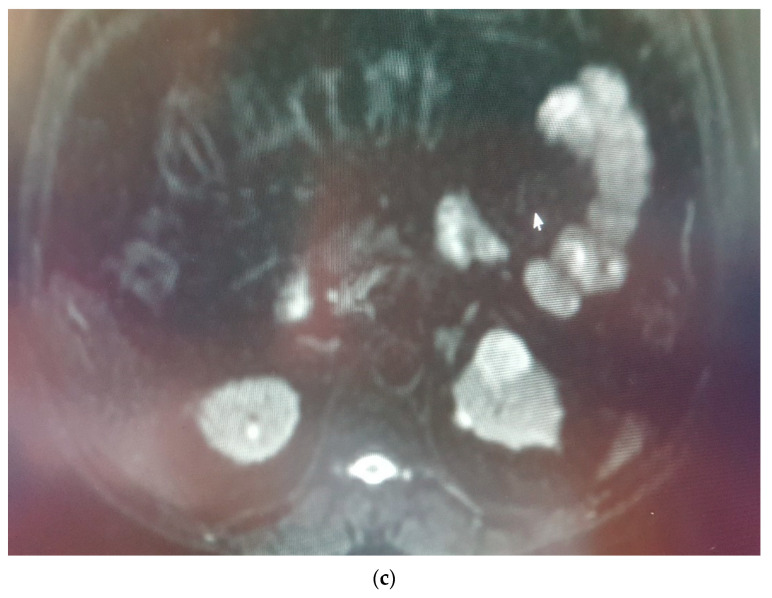
Background and Objectives: Renal Cell Carcinoma (RCC) accounts for 85% and oncocytomas constitute 3-7% of solid renal masses. Oncocytomas can be confused, especially with hypovascular RCC. The purpose of this research was to evaluate the contribution of diffusion-weighted imaging (DWI) and contrast-enhanced MRI sequences in the differential diagnosis of RCC and oncocytoma Materials and Methods: 465 patients with the diagnosis of RCC and 45 patients diagnosed with oncocytoma were retrospectively reviewed between 2009 to 2020. All MRI acquisitions were handled by a 1.5 T device (Achieva, Philips Healthcare, Best, The Netherlands) and all images were evaluated by the consensus of two radiologists with 10-15 years' experience. The SPSS package program version 15.0 software was used for statistical analysis of the study. Chi-square test, Mann-Whitney U test or the Kruskal-Wallis tests were used in the statistical analysis. A receiver operating characteristic (ROC) curve was used to calculate the cut-off values Results: The results were evaluated with a 95% confidence interval and a significance threshold of p < 0.05. ADC values (p < 0.001) and enhancement index (p < 0.01) were significantly lower in the RCC group than the oncocytoma group. Conclusion: DWI might become an alternative technique to the contrast-enhanced MRI in patients with contrast agent nephropathy or with a high risk of nephrogenic systemic fibrosis, calculation of CI of the oncocytoma and RCCs in the contrast-enhanced acquisitions would contribute to the differential diagnosis.
Keywords: ADC; DWI; carcinoma; cell; oncocytoma; renal.
Conflict of interest statement
The authors declare no conflict of interest.
Figures


References
-
- Mytsyk Y., Dutka I., Borys Y., Komnatska I., Shatynska-Mytsyk I., Farooqi A.A., Gazdikova K., Caprida M., Rodrigo L., Kruzliak P. Renal cell carcinoma: Applicability of the apparent coefficient of the diffusion-weighted estimated by MRI for improving their differential diagnosis, histologic subtyping, and differentiation grade. Int. Urol. Nephrol. 2017;49:215–224. doi: 10.1007/s11255-016-1460-3. - DOI - PubMed
MeSH terms
LinkOut - more resources
Full Text Sources
Medical
