. 2022 Feb 19;27(4):1417.
doi: 10.3390/molecules27041417.

PLLA Coating of Active Implants for Dual Drug Release

Affiliations

PLLA Coating of Active Implants for Dual Drug Release

Katharina Wulf et al. Molecules. .

Abstract

Cochlear implants, like other active implants, rely on precise and effective electrical stimulation of the target tissue but become encapsulated by different amounts of fibrous tissue. The current study aimed at the development of a dual drug release from a PLLA coating and from the bulk material to address short-term and long-lasting release of anti-inflammatory drugs. Inner-ear cytocompatibility of drugs was studied in vitro. A PLLA coating (containing diclofenac) of medical-grade silicone (containing 5% dexamethasone) was developed and release profiles were determined. The influence of different coating thicknesses (2.5, 5 and 10 µm) and loadings (10% and 20% diclofenac) on impedances of electrical contacts were measured with and without pulsatile electrical stimulation. Diclofenac can be applied to the inner ear at concentrations of or below 4 × 10⁻⁵ mol/L. Release of dexamethasone from the silicone is diminished by surface coating but not blocked. Addition of 20% diclofenac enhances the dexamethasone release again. All PLLA coatings serve as insulator. This can be overcome by using removable masking on the contacts during the coating process. Dual drug release with different kinetics can be realized by adding drug-loaded coatings to drug-loaded silicone arrays without compromising electrical stimulation.

Keywords: PLLA coating; cochlear implant; diclofenac; dual drug delivery; impedance measurements; spiral ganglion neuron.


Conflict of interest statement

The authors declare no conflict of interest. The funders had no role in the design of the study; in the collection, analyses, or interpretation of data; in the writing of the manuscript, or in the decision to publish the results.

Figures

Figure 1
Figure 1
Survival (a) and neurite length (b) of freshly isolated SGN after addition of different concentrations of DCF, DMS or enalapril. * p < 0.05; *** p < 0.001 against controls. No differences between drugs were detected.
Figure 2
Figure 2
Left: General reaction scheme for the coating of silicone surfaces (Sil) with PLLA via the cross linker GOPS (Sil-GOPS) and the PLLA functionalized with amino groups (Sil-PLLA); Right: representative SEM micrographs of Sil (a); Sil-GOPS (b) and Sil-PLLA (c) surfaces.
Figure 3
Figure 3
FTIR spectra of investigated Sil based samples Sil, Sil-GOPS, Sil-PLLA-NH2, Sil-PLLA in the range of 4000–500 cm−1. The prominent band between 1764–1712 cm−1 corresponds to PLLA signals.
Figure 4
Figure 4
Cumulative in vitro release of incorporated substances from the samples with and without PLLA coating; (a): DMS release from uncoated and coated samples (Ø = 6 mm; DCF: PLLA 5: 95 wt%, N = 3) Release was with p < 0.05 significantly different between all investigated systems after 1 day. (b): DCF release from PLLA coated samples (DCF: PLLA 10: 90 wt% and 20: 80 wt%, N = 3). Release was with p < 0.05 significantly different between days 0 to 71.
Figure 5
Figure 5
Mean (± SEM) impedance values as measured for contacts coated with PLLA at a thickness of 2.5 µm (a), 5 µm (b) or 10 µm (c). All measurements from t = 0 to 24 h were performed with the samples being immersed in 0.9% NaCl. Between all later measurements, left and right contacts were electrically stimulated. N = 5 each; l—left contact of the samples, m—middle, r—right contacts.
Figure 6
Figure 6
Impedance development over time on two contacts, both coated with 5 µm PLLA. Electrical stimulation started right after the 24 h measurement. The dashed horizontal line indicates mean impedance values of uncoated contacts (N = 6).
Figure 7
Figure 7
Mean (± SEM) impedance values as measured for contacts coated with PLLA and loaded with 10% or 20% DCF. All measurements from t = 0 to 24 h were performed with the samples being immersed in 0.9% NaCl. Between all later measurements, contacts were electrically stimulated (N = 8).
Figure 8
Figure 8
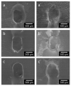
Representative SEM micrographs of platinum contacts after impedance measurements with intact (ac) and damaged (a`c`) PLLA coating of different thicknesses of 2.5 µm (a,a`); 5 µm (b,b`) and 10 µm (c,c`).

References

    1. Anderson I., Baumgartner W.-D., Böheim K., Nahler A., Arnoldner C., Arnolder C., D’Haese P. Telephone use: What benefit do cochlear implant users receive? Int. J. Audiol. 2006;45:446–453. doi: 10.1080/14992020600690969. - DOI - PubMed
    1. Webster M., Webster D.B. Spiral ganglion neuron loss following organ of corti loss: A quantitative study. Brain Res. 1981;212:17–30. doi: 10.1016/0006-8993(81)90028-7. - DOI - PubMed
    1. Scheper V., Paasche G., Miller J.M., Warnecke A., Berkingali N., Lenarz T., Stöver T. Effects of delayed treatment with combined GDNF and continuous electrical stimulation on spiral ganglion cell survival in deafened guinea pigs. J. Neurosci. Res. 2009;87:1389–1399. doi: 10.1002/jnr.21964. - DOI - PubMed
    1. Xu J., Shepherd R.K., Millard R.E., Clark G.M. Chronic electrical stimulation of the auditory nerve at high stimulus rates: A physiological and histopathological study. Hear. Res. 1997;105:1–29. doi: 10.1016/S0378-5955(96)00193-1. - DOI - PubMed
    1. Somdas M.A., Li P.M.M.C., Whiten D.M., Eddington D.K., Nadol J.B. Quantitative evaluation of new bone and fibrous tissue in the cochlea following cochlear implantation in the human. Audiol. Neurootol. 2007;12:277–284. doi: 10.1159/000103208. - DOI - PubMed