

Bruch's-Mimetic Nanofibrous Membranes Functionalized with the Integrin-Binding Peptides as a Promising Approach for Human Retinal Pigment Epithelium Cell Transplantation
- PMID: 35209218
- PMCID: PMC8874486
- DOI: 10.3390/molecules27041429
Bruch's-Mimetic Nanofibrous Membranes Functionalized with the Integrin-Binding Peptides as a Promising Approach for Human Retinal Pigment Epithelium Cell Transplantation
Abstract
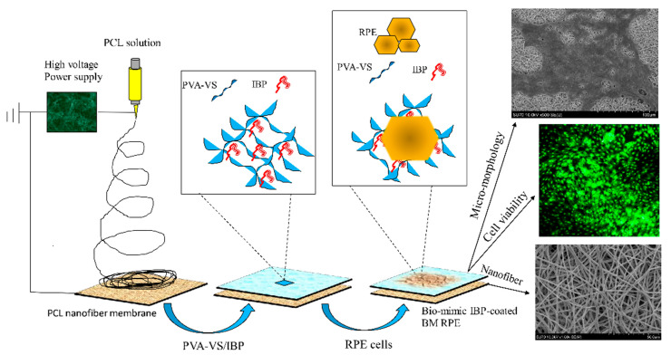
Background: This study aimed to develop an ultrathin nanofibrous membrane able to, firstly, mimic the natural fibrous architecture of human Bruch's membrane (BM) and, secondly, promote survival of retinal pigment epithelial (RPE) cells after surface functionalization of fibrous membranes.
Methods: Integrin-binding peptides (IBPs) that specifically interact with appropriate adhesion receptors on RPEs were immobilized on Bruch's-mimetic membranes to promote coverage of RPEs. Surface morphologies, Fourier-transform infrared spectroscopy spectra, contact angle analysis, Alamar Blue assay, live/dead assay, immunofluorescence staining, and scanning electron microscopy were used to evaluate the outcome.
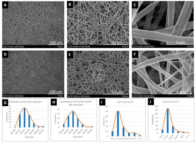
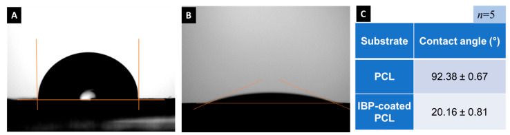
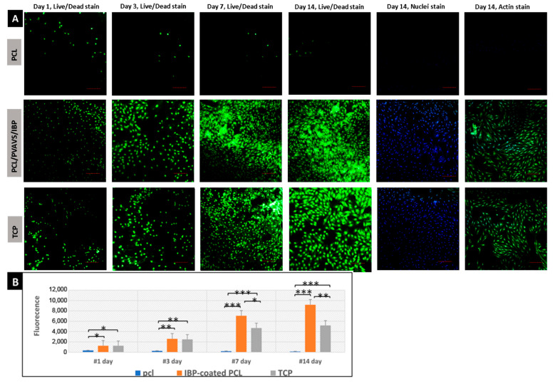
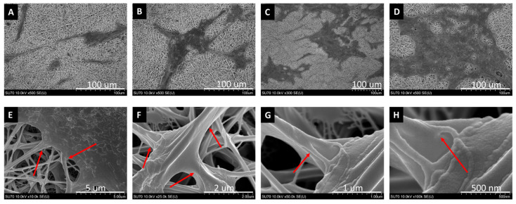
Results: Results showed that coated membranes maintained the original morphology of nanofibers. After coating with IBPs, the water contact angle of the membrane surfaces varied from 92.38 ± 0.67 degrees to 20.16 ± 0.81 degrees. RPE cells seeded on IBP-coated membranes showed the highest viability at all time points (Day 1, p < 0.05; Day 3, p < 0.01; Days 7 and 14, p < 0.001). The proliferation rate of RPE cells on uncoated poly(ε-caprolactone) (PCL) membranes was significantly lower than that of IBP-coated membranes (p < 0.001). SEM images showed a well-organized hexa/polygonal monolayer of RPE cells on IBP-coated membranes. RPE cells proliferated rapidly, contacted, and became confluent. RPE cells formed a tight adhesion with nanofibers under high-magnification SEM. Our findings confirmed that the IBP-coated PCL membrane improved the attachment, proliferation, and viability of RPE cells. In addition, in this study, we used serum-free culture for RPE cells and short IBPs without immunogenicity to prevent graft rejection and immunogenicity during transplantation.
Conclusions: These results indicated that the biomimic BM-IBP-RPE nanofibrous graft might be a new, practicable approach to increase the success rate of RPE cell transplantation.
Keywords: Bruch’s membrane; cell adhesion; electrospinning; immunogenicity; integrin; nanofibers; peptides; retinal pigment epithelial transplantation.
Conflict of interest statement
The authors declare no conflict of interest.
Figures

Similar articles
-
Cytocompatibility of electrospun poly-L-lactic acid membranes for Bruch's membrane regeneration using human embryonic stem cell-derived retinal pigment epithelial cells.J Biomed Mater Res A. 2024 Nov;112(11):1902-1920. doi: 10.1002/jbm.a.37736. Epub 2024 May 10. J Biomed Mater Res A. 2024. PMID: 38726752
-
Primordium of an artificial Bruch's membrane made of nanofibers for engineering of retinal pigment epithelium cell monolayers.Acta Biomater. 2013 Dec;9(12):9414-22. doi: 10.1016/j.actbio.2013.07.029. Epub 2013 Aug 2. Acta Biomater. 2013. PMID: 23917149
-
A novel Bruch's membrane-mimetic electrospun substrate scaffold for human retinal pigment epithelium cells.Biomaterials. 2014 Dec;35(37):9777-9788. doi: 10.1016/j.biomaterials.2014.08.040. Epub 2014 Sep 15. Biomaterials. 2014. PMID: 25220295
-
Biochemical restoration of aged human Bruch's membrane: experimental studies to improve retinal pigment epithelium transplant survival and differentiation.Dev Ophthalmol. 2014;53:133-42. doi: 10.1159/000358531. Epub 2014 Apr 10. Dev Ophthalmol. 2014. PMID: 24732767 Review.
-
Improving RPE adhesion to Bruch's membrane.Eye (Lond). 2009 Oct;23(10):1890-3. doi: 10.1038/eye.2008.411. Epub 2009 Jan 16. Eye (Lond). 2009. PMID: 19151642 Review.
Cited by
-
Advances in Research of Short Peptides.Molecules. 2022 Apr 11;27(8):2446. doi: 10.3390/molecules27082446. Molecules. 2022. PMID: 35458644 Free PMC article.
-
Tissue Engineering of Outer Blood Retina Barrier for Therapeutic Development.Curr Opin Biomed Eng. 2024 Sep;31:100538. doi: 10.1016/j.cobme.2024.100538. Epub 2024 May 13. Curr Opin Biomed Eng. 2024. PMID: 38962280
-
Advances in the engineering of the outer blood-retina barrier: From in-vitro modelling to cellular therapy.Bioact Mater. 2023 Aug 12;31:151-177. doi: 10.1016/j.bioactmat.2023.08.003. eCollection 2024 Jan. Bioact Mater. 2023. PMID: 37637086 Free PMC article. Review.
-
New Advances in Short Peptides: Looking Forward.Molecules. 2022 Jun 6;27(11):3635. doi: 10.3390/molecules27113635. Molecules. 2022. PMID: 35684571 Free PMC article.
References
-
- Bowes Rickman C., LaVail M.M., Anderson E.R., Grimm C., Hollyfield J., Ash J. Retinal Degenerative Diseases: Mechanisms and Experimental Therapy. Springer International Publishing; Cham, Switzerland: 2016. pp. 557–562.
MeSH terms
Substances
LinkOut - more resources
Full Text Sources
