

Machine learning based analysis of stroke lesions on mouse tissue sections
- PMID: 35209753
- PMCID: PMC9274860
- DOI: 10.1177/0271678X221083387
Machine learning based analysis of stroke lesions on mouse tissue sections
Abstract
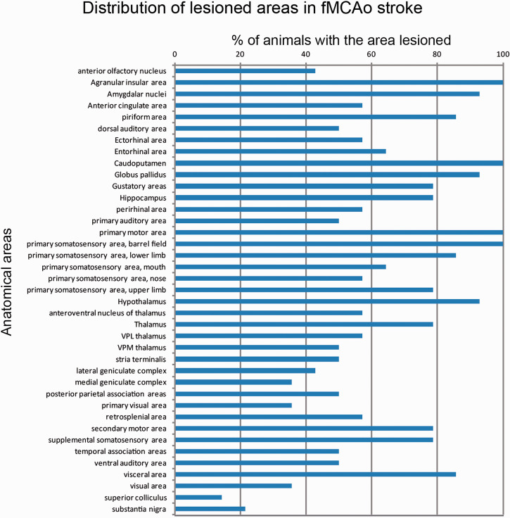
An unbiased, automated and reliable method for analysis of brain lesions in tissue after ischemic stroke is missing. Manual infarct volumetry or by threshold-based semi-automated approaches is laborious, and biased to human error or biased by many false -positive and -negative data, respectively. Thereby, we developed a novel machine learning, atlas-based method for fully automated stroke analysis in mouse brain slices stained with 2% Triphenyltetrazolium-chloride (2% TTC), named "StrokeAnalyst", which runs on a user-friendly graphical interface. StrokeAnalyst registers subject images on a common spatial domain (a novel mouse TTC- brain atlas of 80 average mathematical images), calculates pixel-based, tissue-intensity statistics (z-scores), applies outlier-detection and machine learning (Random-Forest) models to increase accuracy of lesion detection, and produces volumetry data and detailed neuroanatomical information per lesion. We validated StrokeAnalyst in two separate experimental sets using the filament stroke model. StrokeAnalyst detects stroke lesions in a rater-independent and reproducible way, correctly detects hemispheric volumes even in presence of post-stroke edema and significantly minimizes false-positive errors compared to threshold-based approaches (false-positive rate 1.2-2.3%, p < 0.05). It can process scanner-acquired, and even smartphone-captured or pdf-retrieved images. Overall, StrokeAnalyst surpasses all previous TTC-volumetry approaches and increases quality, reproducibility and reliability of stroke detection in relevant preclinical models.
Keywords: Mouse stroke; TTC brain atlas; automated infarct volumetry; lesion analysis; machine learning; neuroanatomical mapping.
Conflict of interest statement
Figures


References
-
- Bederson JB, Pitts LH, Germano SM, et al.. Evaluation of 2,3,5-triphenyltetrazolium chloride as a stain for detection and quantification of experimental cerebral infarction in rats. Stroke 1986; 17: 1304–1308. - PubMed
-
- Lourbopoulos A, Karacostas D, Artemis N, et al.. Effectiveness of a new modified intraluminal suture for temporary middle cerebral artery occlusion in rats of various weight. J Neurosci Methods 2008; 173: 225–234. - PubMed
-
- Tureyen K, Vemuganti R, Sailor KA, et al.. Infarct volume quantification in mouse focal cerebral ischemia: a comparison of triphenyltetrazolium chloride and cresyl violet staining techniques. J Neurosci Methods 2004; 139: 203–207. - PubMed
Publication types
MeSH terms
LinkOut - more resources
Full Text Sources
Medical
