

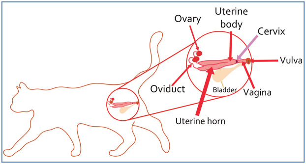
Normal feline reproduction: The queen
- PMID: 35209768
- PMCID: PMC10845401
- DOI: 10.1177/1098612X221079706
Normal feline reproduction: The queen
Abstract
Practical relevance: Cats are seasonally polyestrous, meaning they exhibit multiple estrous cycles within a season, followed by a period of non-cyclicity. Cats cycle when the day length is long but can be induced to cycle year-round with 14 h of continuous artificial lighting. The feline estrous cycle includes the following stages: proestrus, estrus, interestrus and, if ovulation occurs, diestrus. Cats are induced ovulators and ovulate in response to multiple natural matings. Successful breeding in a cattery requires knowledge of the female's reproductive cycle, behavior and management, and often improper management can be the sole cause of infertility.
Aim: The aim of this review is to provide readers with an overview of normal anatomy, cyclicity, management and behavior of the queen. It includes a series of questions veterinarians can ask to obtain a baseline knowledge of the management of the specific breeding set-up.
Evidence base: The information in this article is based on the author's experience, as well as drawing on historical and current literature, and provides the most up-to-date review as possible.
Keywords: Female anatomy; breeding management; cattery management; estrous cycle; puberty.
Conflict of interest statement
The author declared no potential conflicts of interest with respect to the research, authorship and / or publication of this article.
Figures


References
-
- Tsutsui T, Stabenfeldt GH. Biology of ovarian cycles, pregnancy and pseudopregnancy in the domestic cat. J Reprod Fertil Suppl 1993; 47: 29–35. - PubMed
-
- Roberts SJ. Veterinary obstetrics and genital diseases (theriogenology). 3rd ed. Woodstock, VT: Literary Licensing, 1986.
-
- Watson PF, Glover TE. Vaginal anatomy of the domestic cat (Felis catus) in relation to copulation and artificial insemination. J Reprod Fertil Suppl 1993; 47: 355–359. - PubMed
-
- del Campo CH, Ginther oJ. Arteries and veins of uterus and ovaries in dogs and cats. Am J Vet Res 1974; 35: 409–415. - PubMed
-
- Gatel L, Rault dN, Chalvet-Monfray K, et al.. Ultrasonography of the normal reproductive tract of the female domestic cat. Theriogenology 2020; 142: 328–337. - PubMed
Publication types
MeSH terms
LinkOut - more resources
Full Text Sources
Miscellaneous
