. 2022 Feb 24;10(1):26.
doi: 10.1186/s40478-022-01327-6.

Ischemic stroke causes Parkinson's disease-like pathology and symptoms in transgenic mice overexpressing alpha-synuclein

Affiliations

Ischemic stroke causes Parkinson's disease-like pathology and symptoms in transgenic mice overexpressing alpha-synuclein

Stephanie Lohmann et al. Acta Neuropathol Commun. .

Abstract

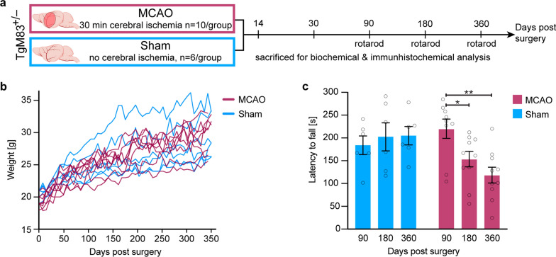
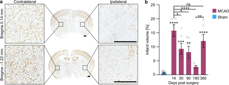
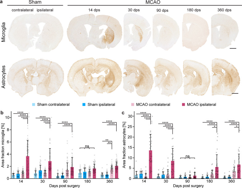
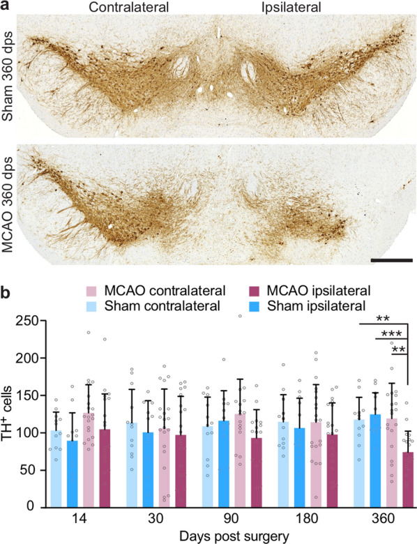
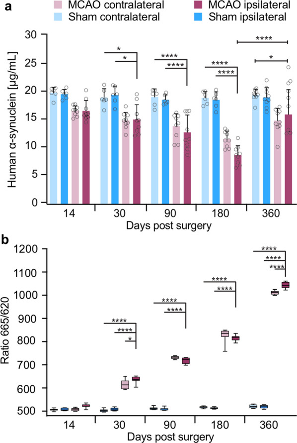
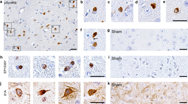
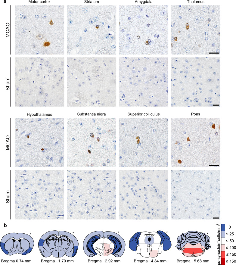
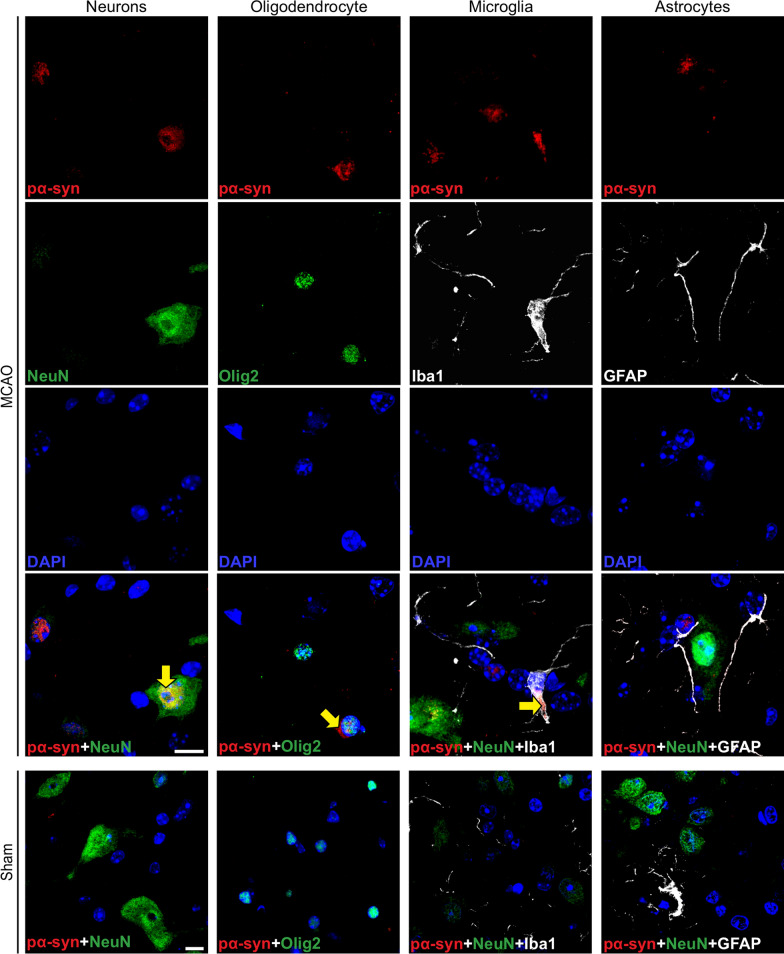
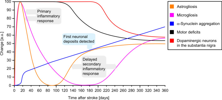
The etiology of Parkinson's disease is poorly understood and is most commonly associated with advancing age, genetic predisposition, or environmental toxins. Epidemiological findings suggest that patients have a higher risk of developing Parkinson's disease after ischemic stroke, but this potential causality lacks mechanistic evidence. We investigated the long-term effects of ischemic stroke on pathogenesis in hemizygous TgM83 mice, which express human α-synuclein with the familial A53T mutation without developing any neuropathology or signs of neurologic disease for more than 600 days. We induced transient focal ischemia by middle cerebral artery occlusion in 2-month-old TgM83+/- mice and monitored their behavior and health status for up to 360 days post surgery. Groups of mice were sacrificed at 14, 30, 90, 180, and 360 days after surgery for neuropathological analysis of their brains. Motor deficits first appeared 6 months after focal ischemia and worsened until 12 months afterward. Immunohistochemical analysis revealed ischemia-induced neuronal loss in the infarct region and astrogliosis and microgliosis indicative of an inflammatory response, which was most pronounced at 14 days post surgery. Infarct volume and inflammation gradually decreased in size and severity until 180 days post surgery. Surprisingly, neuronal loss and inflammation were increased again by 360 days post surgery. These changes were accompanied by a continuous increase in α-synuclein aggregation, its neuronal deposition, and a late loss of dopaminergic neurons in the substantia nigra, which we detected at 360 days post surgery. Control animals that underwent sham surgery without middle cerebral artery occlusion showed no signs of disease or neuropathology. Our results establish a mechanistic link between ischemic stroke and Parkinson's disease and provide an animal model for studying possible interventions.

Keywords: Alpha-synuclein; Ischemia; Ischemic stroke; Parkinson’s disease; Stroke; Synucleinopathy.


Conflict of interest statement

The authors declare that they have no competing interests.

Figures

Fig. 1
Fig. 1
TgM83+/− mice subjected to MCAO gain weight but develop motor deficits within 180 days. TgM83+/− mice expressing the A53T mutant of human α-synuclein were subjected to middle cerebral artery occlusion (MCAO) for 30 min or sham surgery without occlusion of the artery. Groups of treated animals were sacrificed at 14, 30, 90, 180, and 360 days post surgery and neuropathology was analyzed by histology and biochemistry (a). Additionally, the motor behavior of treated animals was tested at 90, 180, and 360 days post surgery on a rotarod treadmill (a). TgM83+/− mice subjected to MCAO (n = 8, red lines) as well as sham-treated animals (n = 6, blue lines) gained body weight until the end of the experiment at 360 days post surgery (b). MCAO- and sham-treated mice were tested for motor impairment using a rotarod at 90, 180, and 360 days post surgery. TgM83+/− mice that had been subjected to MCAO (red) showed significantly reduced motor skills at 180 and 360 days post surgery relative to 90 days post surgery (c). In contrast, sham-treated control animals (blue) did not develop any motor deficits. Six to ten animals were analyzed per group. The data represents the mean latency to fall in seconds ± standard error of the mean. P values were computed using two-way ANOVA followed by Tukey’s post-hoc test (P: * < 0.05, ** < 0.01)
Fig. 2
Fig. 2
TgM83+/− mice show neuronal loss in the ipsilateral brain hemisphere after MCAO. Immunohistochemical staining for the neuronal marker NeuN showed an ipsilateral loss of neurons in ischemic brains of TgM83+/− mice after MCAO (a). Quantification of brain tissue sections ranging from bregma 0.14 mm to −1.22 mm of mice subjected to MCAO revealed significant ipsilateral neuronal degeneration in the ischemic core and peri-infarct region at 14, 30, 90, 180, and 360 days post surgery relative to sham-treated animals (b). The infarct size was calculated in relation to the contralateral brain hemisphere to minimize the error caused by edema. Two coronal brain sections of three MCAO-treated animals were analyzed for each time point and two coronal brain sections of eight sham-treated animals for all time points together. The data represents the mean infarct volume in percent ± standard error of the mean. P values were computed using one-way ANOVA followed by Tukey’s post-hoc test (P: **** < 0.0001, *** < 0.001, ** < 0.01). The scale bar represents 0.5 mm
Fig. 3
Fig. 3
TgM83+/− mice display early and late inflammatory responses in the brain after MCAO. Microglia and astrocytes in coronal brain tissue sections of MCAO- and sham-treated TgM83+/− mice were stained with antibodies to Iba1 or to GFAP, respectively (a). Quantification of the stainings revealed that microgliosis (b) and astrogliosis (c) were significantly induced at 14 days post surgery within the stroke lesion area of mice subjected to MCAO (dark red) when compared to the contralateral hemisphere (light red), or the ipsi- (dark blue) and contralateral (light blue) brain hemispheres of sham-treated animals. Beginning at 30 days post surgery we measured a continuous decline in the level of inflammation, which was reduced to levels observed in sham-treated animals at 90 days post surgery for astrogliosis and later at 180 days post surgery for microgliosis. Following its initial clearance, the inflammatory reaction, surprisingly, began to resurface again in the brains of MCAO-treated animals. We measured a continuous rise of astrogliosis from 90 to 360 days post surgery in the ipsilateral brain hemisphere of MCAO-treated animals. At 360 days post surgery also microgliosis was significantly induced again in the ipsilateral and, importantly, also in the contralateral brain hemisphere of MCAO-treated animals. A continuous increase of astrogliosis was also measured in the contralateral brain hemisphere of MCAO-treated animals at 180 and 360 days post surgery but did not reach significance. The data represent the mean area fraction in percent ± standard deviation. Six to twelve coronal brain sections of three to five animals were analyzed per group at 14, 30, 180, and 360 days post surgery. P values were computed using two-way ANOVA followed by Tukey’s post-hoc test (P: * < 0.05, **** < 0.0001). Scale bars represent 1.0 mm
Fig. 4
Fig. 4
MCAO-treated TgM83+/− mice display a late loss of dopaminergic neurons in the substantia nigra. Immunhistochemical analysis of the substantia nigra of MCAO- and sham-treated TgM83+/− mice with an antibody against TH showed an ipsilateral loss of dopaminergic neurons 360 days after MCAO (a). Quantification of TH-positive cells revealed a significant loss of dopaminergic neurons on the ipsilateral side of the substantia nigra of TgM83+/− mice after MCAO-treatment (dark red) relative to the contralateral side (light red) or to sham-treated control animals (dark and light blue) at 360 days post surgery (b). The data represent the mean count of TH-positive cells per coronal section of the substantia nigra ± standard deviation. Four coronal brain sections of three to five animals were analyzed per group 14, 30, 90, 180, and 360 days post surgery. P values were computed using two-way ANOVA followed by Tukey’s post-hoc test (P: ** < 0.01, *** < 0.001). The scale bar represents 0.5 mm
Fig. 5
Fig. 5
MCAO induces a continuous long-term increase of aggregated α-synuclein species in the CNS of TgM83+/− mice. Quantification of the amount of human α-synuclein protein by ELISA in brain homogenates of TgM83+/− mice subjected to MCAO showed a continuous decline in the ipsi- (dark red) and contralateral (light red) brain hemisphere up to 180 days post surgery, after which α-synuclein levels slightly rose again but did not reach levels detected in the brains of animals at 360 days post sham surgery (a). No changes in α-synuclein protein levels were observed in the ipsi- (dark blue) or contralateral (light blue) brain hemisphere of animals after sham surgery. Quantification of aggregated α-synuclein species by FRET analysis in brain homogenates of TgM83+/− mice revealed a steady increase in the ipsi- (dark red) and contralateral (light red) brain hemisphere beginning 30 days post surgery (b). In contrast, α-synuclein did not aggregate in the brains of TgM83+/− mice after sham surgery. Both brain hemispheres of three animals each were analyzed per group at 14, 30, 90, 180, and 360 days post surgery. The ELISA data represents the mean ± standard deviation. The FRET data are shown as boxplots. P values were computed using two-way ANOVA followed by Tukey’s post-hoc test (P: * < 0.05, **** < 0.0001)
Fig. 6
Fig. 6
Diseased TgM83+/− mice display various types of pathologic α-synuclein deposits in the CNS. Immunohistochemical staining of coronal brain tissues sections of diseased TgM83+/− mice revealed abundant deposits of phosphorylated α-synuclein throughout the brain at 360 days after stroke using the pSyn#64 (a–f), EP1536Y (h), and 81A (j) antibodies that recognize phosphorylated α-synuclein at Ser129, which is associated with the accumulation of pathologic α-synuclein. Deposits of phosphorylated α-synuclein were mostly cytosolic (b–d), sometimes perinuclear (e), or dendritic (arrow) (f). In contrast, sham-treated mice did not show any deposits of phosphorylated α-synuclein with any of the three antibodies used (g, i, k). Staining with the 81A antibody resulted in a higher background due to a known cross reactivity of this antibody with phosphorylated neurofilament subunit L (j, k). The scale bars represent 20 µm
Fig. 7
Fig. 7
Diseased TgM83+/− mice harbor deposits of pathologic α-synuclein in various brain regions. To reveal deposits of pathologic α-synuclein, coronal tissue sections of different brain regions of diseased TgM83+/− mice collected at 360 days after stroke were stained with the pSyn#64 antibody against α-synuclein phosphorylated at serine 129. Diseased animals that had been subjected to stroke but not sham-treated animals showed cellular deposits of pathologic α-synuclein throughout the cerebrum, including the motor cortex, the striatum, the amygdala, the thalamus, the hypothalamus, the substantia nigra, the superior colliculus, and the pons (a). Deposits of pathologic α-synuclein in the brains of diseased TgM83+/− mice at 360 days after MCAO were quantified as phosphorylated α-synuclein-positive cells per square millimeter from four diseased animals per brain region (Additional file 4), and the data were summarized in heat maps spanning five rostral-to-caudal brain areas (b). Asterisks indicate the stroke side. Scale bars represent 20 µm
Fig. 8
Fig. 8
Pathologic α-synuclein colocalizes with neurons, oligodendrocytes, and microglia in the CNS of diseased TgM83+/− mice. Immunofluorescence staining of tissue sections of the brain stem show that phosphorylated α-synuclein (pSyn#64, red) colocalizes with neurons (NeuN, green) as well as with oligodendrocytes (Olig2, green) in mice 360 days after MCAO as indicated by yellow arrows. Deposits of phosphorylated α-synuclein (pSyn#64, red) were additionally seen to colocalize with microglia (Iba1, white) but not with astrocytes (GFAP, white) as indicated by yellow arrows. Neurons, oligodendrocytes, microglia, or astrocytes of mice that underwent sham-surgery, for which only merged images are shown, did not accumulate any phosphorylated α-synuclein deposits. Nuclear staining with DAPI is shown in blue. The scale bar represents 10 µm
Fig. 9
Fig. 9
Timeline of pathogenesis in the CNS of TgM83+/− mice after ischemic stroke. Ischemia-induced astrogliosis (orange line) and microgliosis (pink line) both peaked at 14 days after the initial insult, which is also when α-synuclein aggregation (blue line) steadily began to increase based on FRET analysis. Astrogliosis and microgliosis returned close to their pre-ischemic levels by 90 and 180 days post ischemia, respectively, while α-synuclein aggregation continued to increase. Possibly due to the gradually increasing load and deposition of aggregated α-synuclein in the brain, a second inflammatory process set in with a renewed increase in astrogliosis that was slowly followed by microgliosis. By 180 days post ischemia, when cellular deposits of pathologic α-synuclein were first detected, animals also showed first symptoms of motor disease (black line), which worsened over time. By 360 days post ischemia a loss of dopaminergic neurons was detected in the ipsilateral side of the substantia nigra (red line)

References

    1. Dement A. Alzheimer's disease facts and figures. Alzheimer's Dementia. 2020;16:391–460. doi: 10.1002/alz.12068. - DOI
    1. Alvarez-Castelao B, Goethals M, Vandekerckhove J, Castano JG. Mechanism of cleavage of alpha-synuclein by the 20S proteasome and modulation of its degradation by the RedOx state of the N-terminal methionines. Biochem Biophys Acta. 2014;1843:352–365. doi: 10.1016/j.bbamcr.2013.11.018. - DOI - PubMed
    1. Ascherio A, Schwarzschild MA. The epidemiology of Parkinson's disease: risk factors and prevention. Lancet Neurol. 2016;15:1257–1272. doi: 10.1016/S1474-4422(16)30230-7. - DOI - PubMed
    1. Baba M, Nakajo S, Tu PH, Tomita T, Nakaya K, Lee VM, Trojanowski JQ, Iwatsubo T. Aggregation of alpha-synuclein in Lewy bodies of sporadic Parkinson's disease and dementia with Lewy bodies. Am J Pathol. 1998;152:879–884. - PMC - PubMed
    1. Barbour R, Kling K, Anderson JP, Banducci K, Cole T, Diep L, Fox M, Goldstein JM, Soriano F, Seubert P, et al. Red blood cells are the major source of alpha-synuclein in blood. Neurodegener Dis. 2008;5:55–59. doi: 10.1159/000112832. - DOI - PubMed

Publication types

Substances