Case Reports
. 2022 Feb 24;15(2):e244732.
doi: 10.1136/bcr-2021-244732.

Abdominal Kikuchi-Fujimoto lymphadenopathy: an uncommon presentation of a rare disease

Affiliations
Case Reports

Abdominal Kikuchi-Fujimoto lymphadenopathy: an uncommon presentation of a rare disease

Marco Fiori et al. BMJ Case Rep. .

Abstract

A 34-year-old man presented to our hospital with a 5-day history of progressive abdominal pain and fever. A CT scan identified extensive mesenteric lymphadenopathy. Initial diagnostic tests were inconclusive. Abdominal lymph node biopsy showed histiocytic necrotising lymphadenitis, compatible with Kikuchi-Fujimoto disease (KFD). This benign and self-limiting disease generally resolves following supportive treatment. In this case, remission occurred within 3 weeks of initial presentation. KFD is a very uncommon cause of lymphadenopathy, and selective mesenteric involvement is rare. Definitive diagnosis often requires lymph node biopsy. It is important to exclude more common and serious differential diagnoses associated with mesenteric lymphadenopathy, while maintaining a minimally invasive diagnostic approach, before progressing to nodal biopsy.

Keywords: haematology (incl blood transfusion); immunology; pathology.


Conflict of interest statement

Competing interests: None declared.

Figures
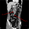
Figure 1
Figure 1
Multiple enlarged lymph nodes (*) along the mesenteric vessels (maximum diameter up to 22×8 mm) associated with thickening of the mesenteric adipose tissue (**).
Figure 2
Figure 2
Histological examination of histiocytic necrotising lymphadenitis (Kikuchi-Fujimoto disease). H&E staining (top, H&E, ×200) shows subverted architecture due to expansion of the interfollicular and paracortical zones, with necrosis rich in karyorrhectic nuclear debris, without significant polymorphonuclear leucocytes component. Immunohistochemical analyses (bottom, CD68 staining, ×200) show various type of macrophages, including plasmacytoid dendritic cells.

References

    1. Perry AM, Choi SM. Kikuchi-Fujimoto disease: a review. Arch Pathol Lab Med 2018;142:1341–6. 10.5858/arpa.2018-0219-RA - DOI - PubMed
    1. Ahmed Z, Quadir H, Hakobyan K, et al. . Kikuchi-Fujimoto disease: a rare cause of cervical lymphadenopathy. Cureus 2021;5. 10.7759/cureus.17021 - DOI - PMC - PubMed
    1. Huang J, Zheng J-X, Yang Y, et al. . Necrotizing lymphadenitis. Z Rheumatol 2021;80:274–82. 10.1007/s00393-020-00929-6 - DOI - PubMed
    1. Singh JM, Shermetaro CB. Kikuchi-Fujimoto disease in Michigan: a rare case report and review of the literature. Clin Med Insights Ear Nose Throat 2019;12:117955061982868. 10.1177/1179550619828680 - DOI - PMC - PubMed
    1. Helbling R, Conficconi E, Wyttenbach M, et al. . Acute Nonspecific Mesenteric Lymphadenitis: More Than “No Need for Surgery”. Biomed Res Int 2017;2017:1–4. 10.1155/2017/9784565 - DOI - PMC - PubMed

Publication types

MeSH terms