. 2022 Mar 1;119(9):e2112840119.
doi: 10.1073/pnas.2112840119.

An immune-sympathetic neuron communication axis guides adipose tissue browning in cancer-associated cachexia

Affiliations

An immune-sympathetic neuron communication axis guides adipose tissue browning in cancer-associated cachexia

Hao Xie et al. Proc Natl Acad Sci U S A. .

Abstract

Cancer-associated cachexia (CAC) is a hypermetabolic syndrome characterized by unintended weight loss due to the atrophy of adipose tissue and skeletal muscle. A phenotypic switch from white to beige adipocytes, a phenomenon called browning, accelerates CAC by increasing the dissipation of energy as heat. Addressing the mechanisms of white adipose tissue (WAT) browning in CAC, we now show that cachexigenic tumors activate type 2 immunity in cachectic WAT, generating a neuroprotective environment that increases peripheral sympathetic activity. Increased sympathetic activation, in turn, results in increased neuronal catecholamine synthesis and secretion, β-adrenergic activation of adipocytes, and induction of WAT browning. Two genetic mouse models validated this progression of events. 1) Interleukin-4 receptor deficiency impeded the alternative activation of macrophages, reduced sympathetic activity, and restrained WAT browning, and 2) reduced catecholamine synthesis in peripheral dopamine β-hydroxylase (DBH)-deficient mice prevented cancer-induced WAT browning and adipose atrophy. Targeting the intraadipose macrophage-sympathetic neuron cross-talk represents a promising therapeutic approach to ameliorate cachexia in cancer patients.

Keywords: adipose tissue; browning; cancer cachexia; immunometabolism; macrophage.


Conflict of interest statement

The authors declare no competing interest.

Figures
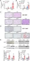
Fig. 1.
Fig. 1.
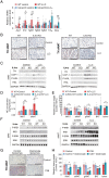
CAC is associated with increased plasma IL-6 and PTHrP concentrations and WAT browning. (AE) Mice bearing LLC, C26, or C26nc tumors were euthanized 14 to 16 d after tumor cell inoculation. (A and B) PTHrP and IL-6 plasma concentrations were determined in LLC (A), C26, and C26nc (B) tumor-bearing mice using ELISA (n = 4 to 5 per group). (C and D) Representative iWAT sections of LLC, C26, and C26nc tumor-bearing mice stained with hematoxylin and eosin (H&E; C) or immunolabeled for UCP-1 (D). (Scale bars: 200 µm.) (E) Western blotting analysis to detect uncoupling protein-1 (UCP-1) protein expression in iWAT and aWAT of LLC, C26, and C26nc tumor-bearing mice. Glycerinaldehyde-3-phosphate-dehydrogenase (GAPDH) was used as loading control. The protein contents of UCP-1 and GAPDH were quantified by densitometric analysis. Data are presented as means ± SD. Significance was determined by unpaired two-tailed Student’s t test (A and E) or a one-way ANOVA followed by Tukey’s post hoc analysis. *P ≤ 0.05; **P ≤ 0.01.
Fig. 2.
Fig. 2.
IL-6 and PTHrP do not directly stimulate thermogenic gene expression or lipolysis in white and brown adipocyte cell lines. (A and B) qRT-PCR analysis was used to detect mRNA levels of thermogenic marker genes in differentiated 3T3-L1 (white adipocytes; A) and iBACs (brown adipocytes; B) treated with 10 ng/mL PTHrP, 20 ng/mL IL-6, 20 ng/mL IL-6/37.14 ng/mL (equimolar to IL-6) sIL-6ra, 10 µM forskolin, or vehicle for 8 h. (C and D) Glycerol release from differentiated 3T3-L1 (C) and iBACs (D) treated with 40 ng/mL PTHrP, 20 ng/mL IL-6, 20 ng/mL IL-6/37.14 ng/mL (equimolar to IL-6) sIL-6ra, 10 µM forskolin, or vehicle and 2% FA-free BSA for 8 h. Data are presented as means ± SD. Significance was determined by one-way ANOVA followed by Tukey’s post hoc analysis. *P ≤ 0.05; **P ≤ 0.01. (E) Legend for A–D.
Fig. 3.
Fig. 3.
CAC is associated with increased catecholamine concentrations and elevated tyrosine hydroxylase (TH) expression in adipose tissue. (AD) Mice bearing LLC, C26, or C26nc tumors were euthanized 14 to 16 d after tumor cell inoculation. (A and B) Noradrenaline (NE) contents in iWAT and BAT sections (A) as well as in plasma (B) of LLC, C26, and C26nc tumor-bearing mice were determined by ELISA (n = 3 to 4 per group). (C) Western blotting analysis to detect TH protein expression in iWAT and aWAT of LLC, C26, and C26nc tumor-bearing mice. Glycerinaldehyde-3-phosphat-dehydrogenase (GAPDH) was used as loading control. The protein contents of TH or GAPDH were quantified by densitometric analysis. (D) Representative iWAT sections of LLC, C26, and C26nc tumor-bearing mice immunolabeled for TH. (Scale bars: 200 µm.) (E and F) Representative three-dimensional projections of whole iWAT depots from (E) LLC, (F) C26, and C26nc tumor-bearing mice immunolabeled for TH and imaged at 1.26× magnification on a light-sheet microscope. (Scale bars: 5,000 µm.) (G) For quantification, randomly selected cubes were isolated from adipose tissue blocks that were derived from identical positions of whole adipose tissue depots of three to six mice per group. Neuron density in each cube was calculated as the ratio of total neurite length per regional volume. The mean of five cubes from each tissue block was calculated and represents one data point in the graph. Data are presented as means ± SD. Significance was determined by unpaired two-tailed Student’s t test or a one-way ANOVA followed by Tukey’s post hoc analysis. *P ≤ 0.05; **P ≤ 0.01.
Fig. 4.
Fig. 4.
Deletion of dopamine β-hydroxylase (DBH) in peripheral tissues results in catecholamine depletion, reduced lipolysis, and impaired browning in adipose tissue of cachexigenic tumor-bearing mice. (AC) LLC tumor-bearing mice were euthanized 14 to 16 d after tumor cell inoculation. (A) Tissue weights of DBHΔper LLC tumor-bearing mice relative to WT control animals (n = 9 to 10 per group; results of two independent experiments were combined). (B) NE contents of iWAT and aWAT of WT and DBHΔper LLC tumor-bearing mice and the respective control mice were determined by ELISA (n = 4 per group). (C) Western blotting analysis to detect DBH, tyrosine hydroxylase (TH), uncoupling protein-1 (UCP-1), phosphorylated (P)-HSL (Ser660), and HSL protein contents in iWAT and aWAT of LLC tumor-bearing and control WT and DBHΔper mice. Glycerinaldehyde-3-phosphate-dehydrogenase (GAPDH) was used as the loading control. Data are presented as means ± SD. Significance was determined by two-way ANOVA followed by Tukey’s post hoc analysis. *P ≤ 0.05; **P ≤ 0.01.
Fig. 5.
Fig. 5.
Alternatively activated macrophages promote sympathetic neurite outgrowth in CAC. (AD) Mice bearing LLC, C26, or C26nc tumors were euthanized 14 to 16 d after tumor cell inoculation. (A and B) qRT-PCR analysis was used to detect mRNA levels of Ngf (A) and Gap-43 (B) in iWAT (n = 5 to 10 per group; results from two independent experiments were combined). (C and D) Representative three-dimensional projections of iWAT depots from LLC (C), C26, and C26nc (D) tumor-bearing mice immunolabeled for GAP-43 and imaged at 1.26× magnification using a light-sheet microscope. For quantification, randomly selected cubes were isolated from adipose tissue blocks that were derived from identical positions of whole adipose tissue depots of three to six mice per group. Neuron density in each cube was calculated as the ratio of total neurite length per regional volume. The mean of five cubes from each tissue block was calculated and represents one data point in the graph. (Scale bars: 5,000 µm.) (E and F) Representative images of cultured primary sympathetic neurons grown in the presence or absence of 10 ng/mL PTHrP, 20 ng/mL IL-6, or 20 ng/mL IL-6/37.14 ng/mL (equimolar to IL-6) sIL-6ra (E) or cocultured with primary macrophages in the presence and absence of IL-4 and 2% plasma from control or LLC tumor-bearing (CAC) mice (F), immunolabeled for TH, and imaged by confocal fluorescence microscopy. Neurite length was determined using the Neuron J plug-in of ImageJ (n = 9 per condition). (Scale bars: 100 µm.) (G) Primary macrophages were incubated in the presence and absence of IL-4 and 2% plasma from control or LLC tumor-bearing (CAC) mice for 24 h and analyzed for neurotrophic gene expression using qRT-PCR (n = 3 per condition). Data are presented as means ± SD. Significance was determined by unpaired two-tailed Student’s t test or a one-way ANOVA (A and E) or by a two-way ANOVA followed by Tukey’s post hoc analysis (F and G). *P ≤ 0.05; **P ≤ 0.01.
Fig. 6.
Fig. 6.
IL-4ra deficiency or local macrophage depletion prevents CAC-induced catecholamine synthesis and browning of adipose tissue. (AF) WT and IL4ra-knock out (KO) mice bearing LLC tumors were euthanized 14 to 16 d after tumor cell inoculation. (A) qRT-PCR analysis to detect mRNA levels of type 1 and type 2 immune cell marker genes in iWAT (n = 4 to 5 per group). (B) Representative iWAT and aWAT sections immunolabeled for tyrosine hydroxylase (TH). (Scale bars: 200 µm.) (C) Western blotting analysis to detect TH, uncoupling protein-1 (UCP-1), phosphorylated (P) (Ser660) hormone sensitive lipase (P-HSL), and HSL protein contents in iWAT and aWAT. Glycerinaldehyde-3-phosphat-dehydrogenase (GAPDH) was used as loading control. (D) Norepinephrine (NE) contents in iWAT and aWAT were determined by ELISA (n = 4 per group). (E) Tissue weights of WT and IL4ra-KO LLC tumor-bearing mice relative to tissue weights of control animals (n = 10 per group; results of two independent experiments were combined). (F) Western blotting analysis to detect phosphorylated (P) (Ser724)-inositol-requiring protein-1 (P-IRE1), IRE1, phosphorylated (P) (Thr202/Tyr204)-extracellular signal–regulated kinase (P-ERK), and ERK protein expression in iWAT and aWAT depots. (G and H) C26 tumor-bearing mice and control mice were subcutaneously injected with control/clodronate liposomes every 2 d and euthanized 14 to 16 d after tumor cell inoculation. (G) Representative iWAT sections of control and C26 tumor-bearing mice treated with clodronate/control liposomes immunolabeled for TH. (Scale bars: 200 µm.) (H) Tissue weights of control/clodronate liposomes–treated C26 tumor-bearing mice relative to tissue weights of the respective control animals (n = 4 to 5 per group). Data are presented as means ± SD. Significance was determined by two-way ANOVA followed by Tukey’s post hoc analysis. *P ≤ 0.05; **P ≤ 0.01.
Fig. 7.
Fig. 7.
Mechanisms of WAT browning in CAC. A chronic inflammatory, hypercatabolic state causes metabolic adaptations in WAT, summarized in the term browning, which contribute to WAT atrophy. We demonstrate that cachexia-associated proinflammatory factors, like IL-6 and PTHrP, do not directly induce a catabolic switch in adipocytes but likely activate an intraadipose macrophage-sympathetic neuron cross-talk that orchestrates browning of WAT. The proinflammatory environment, generated by the cachexigenic tumor, causes the infiltration and IL-4–dependent alternative activation of macrophages (blue) in WAT, whereas the amount of proinflammatory macrophages (red) was unaffected. Alternatively activated macrophages secrete NGF, BDNF, and polyamines to generate a neuroprotective environment that allows for an increased GAP43-conducted outgrowth and activity of sympathetic neuronal projections. Enhanced sympathetic activity results in increased neuronal catecholamine secretion, β-adrenergic activation of adipocytes, and WAT browning.

References

    1. Porporato P. E., Understanding cachexia as a cancer metabolism syndrome. Oncogenesis 5, e200–e210 (2016). - PMC - PubMed
    1. von Haehling S., Anker S. D., Cachexia as a major underestimated and unmet medical need: Facts and numbers. J. Cachexia Sarcopenia Muscle 1, 1–5 (2010). - PMC - PubMed
    1. Petruzzelli M., et al. , A switch from white to brown fat increases energy expenditure in cancer-associated cachexia. Cell Metab. 20, 433–447 (2014). - PubMed
    1. Kir S., et al. , Tumour-derived PTH-related protein triggers adipose tissue browning and cancer cachexia. Nature 513, 100–104 (2014). - PMC - PubMed
    1. Arora G. K., et al. , Cachexia-associated adipose loss induced by tumor-secreted leukemia inhibitory factor is counterbalanced by decreased leptin. JCI Insight 3, e121221 (2018). - PMC - PubMed

Substances