. 2022 Mar 1;119(9):e2115316119.
doi: 10.1073/pnas.2115316119.

HTLV-1 activates YAP via NF-κB/p65 to promote oncogenesis

Affiliations

HTLV-1 activates YAP via NF-κB/p65 to promote oncogenesis

Tiejun Zhao et al. Proc Natl Acad Sci U S A. .

Abstract

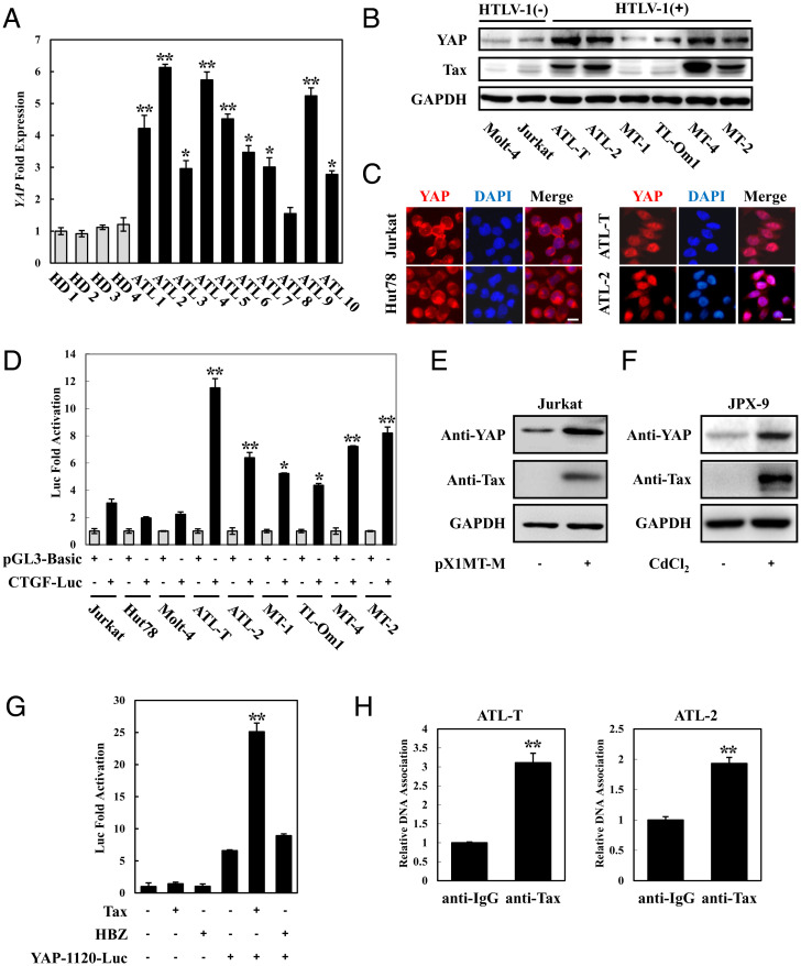
Adult T-cell leukemia/lymphoma (ATL) is an aggressive malignancy caused by human T-cell leukemia virus type 1 (HTLV-1) infection. HTLV-1 exerts its oncogenic functions by interacting with signaling pathways involved in cell proliferation and transformation. Dysregulation of the Hippo/YAP pathway is associated with multiple cancers, including virus-induced malignancies. In the present study, we observe that expression of YAP, which is the key effector of Hippo signaling, is elevated in ATL cells by the action of the HTLV-1 Tax protein. YAP transcriptional activity is remarkably enhanced in HTLV-1-infected cells and ATL patients. In addition, Tax activates the YAP protein via a mechanism involving the NF-κB/p65 pathway. As a mechanism for this cross talk between the Hippo and NF-κB pathways, we found that p65 abrogates the interaction between YAP and LATS1, leading to suppression of YAP phosphorylation, inhibition of ubiquitination-dependent degradation of YAP, and YAP nuclear accumulation. Finally, knockdown of YAP suppresses the proliferation of ATL cells in vitro and tumor formation in ATL-engrafted mice. Taken together, our results suggest that p65-induced YAP activation is essential for ATL pathogenesis and implicate YAP as a potential therapeutic target for ATL treatment.

Keywords: ATL; HTLV-1; Tax; YAP; p65.


Conflict of interest statement

The authors declare no competing interest.

Figures

Fig. 1.
Fig. 1.
YAP is up-regulated and activated in ATL. (A) High expression of YAP in ATL patients. CD4-positive cells were isolated from PBMCs of healthy donors and ATL patients. qPCR was performed to analyze the expression of YAP. HD 1 to 4 indicates healthy donors, and ATL 1 to 10 indicates ATL patients. (B) YAP protein was up-regulated in ATL cell lines. Total protein was extracted from HTLV-1–negative and HTLV-1–positive cell lines and subjected to Western blot using YAP and Tax antibodies. (C) YAP localized in the nucleus of ATL cell lines. YAP subcellular localization was determined by immunofluorescence staining for YAP (red) and 4',6-diamidino-2-phenylindole (DAPI) for DNA (blue). (Scale bar, 10 μm.) (D) The Hippo pathway was suppressed in ATL cells. Cells were cotransfected with pGL3-Basic or CTGF-Luc together with phRL-TK. Forty-eight hours after transfection, cells were harvested and analyzed for luciferase activity. (E) HTLV-1 induced YAP expression. Jurkat cells were transfected with an infectious molecular clone of HTLV-1 (pX1MT-M). Forty-eight hours posttransfection, the expression of YAP was analyzed by immunoblotting. (F) YAP protein was up-regulated in Tax-expressing JPX-9 cells. JPX-9 cells were treated with 30 μmol/L of CdCl2 for 18 h. After incubation, the expression of Tax and YAP protein was examined by immunoblot analysis. (G) Tax enhanced YAP promoter activity. Jurkat cells were transfected with YAP reporter vector (YAP-1120-Luc) and Tax or HBZ expression plasmid. Luciferase activity was measured 48 h after transfection. (H) ChIP analysis of the association of endogenous Tax and YAP promoters in ATL cells (ATL-T and ATL-2). The statistical analyses were performed by unpaired two-tailed Student's t test. *P < 0.05, **P < 0.01.
Fig. 2.
Fig. 2.
Tax activates YAP via p65. (A) Tax stimulated YAP transcriptional activation. Jurkat cells were cotransfected with 8×GTIIC–Luc (0.5 μg), phRL-TK (20 ng), and pCG-Tax (1 or 3 μg) with or without pCMV-FLAG-YAP (0.1 μg). Forty-eight hours after transfection, the cells were harvested and analyzed for luciferase activity. (B) HBZ had no effect on YAP activity. Jurkat cells were cotransfected with 8×GTIIC–Luc, phRL-TK, pCMV-FLAG-YAP, and pME18Sneo-HBZ. After 48 h, a dual-luciferase reporter assay was performed. (C) Analysis of Tax mutants for their effect on YAP-induced transcriptional activation. Jurkat cells were cotransfected with 8×GTIIC–Luc, phRL-TK, pCMV-FLAG-YAP, and pCG-Tax or its mutants. Luciferase activity was measured 48 h after transfection. (D) Tax activated YAP transcription depending on NF-κB. Jurkat cells were cotransfected with 8×GTIIC–Luc, phRL-TK, pCMV-FLAG-YAP, pCG-Tax, and expression plasmid for IκB (S32A/S36A) or ACREB. At 48 h after transfection, cell lysates were subjected to luciferase assay. (E) p65 enhanced YAP activity. Jurkat cells were cotransfected with 8×GTIIC–Luc, phRL-TK, pCMV-FLAG-YAP, and pSG-p65. At 48 h after transfection, the cells were harvested, and luciferase activity was measured. The statistical analyses were performed by unpaired two-tailed Student's t test. *P < 0.05, **P < 0.01.
Fig. 3.
Fig. 3.
p65 interacts with YAP to enhance YAP activity. (A) p65 interacted with YAP but not LAST1. The 293T cells were transfected with the indicated expression vectors. Cell lysates were subjected to immunoprecipitation (IP) using anti-FLAG followed by immunoblotting (IB) using anti-HA. The expression levels of YAP, p65, and LATS1 were detected. (B) YAP interacted with p65 endogenously. Whole-cell lysate of ATL-T cells was subjected to immunoprecipitation with anti-YAP or control IgG, and immunoprecipitates were probed with anti-p65 antibody. The band of p65 which coimmunoprecipitated with YAP is indicated with an asterisk. (C) YAP colocalizated with p65 in the cell nucleus. The 293T cells were transfected with pCMV-FLAG-YAP together with (vvii) or without (i and ii) pcDNA-MycHis-p65. p65 was detected using anti-MYC Cy3 antibody (iii and vi). YAP was detected using anti–FLAG-biotin and secondary Streptavidin-Alexa 488 antibody (i and v). The overlay of YAP and p65 is shown (vii). DAPI (4,6 diamidino-2-phenylindole) was used to counterstain the nucleus (ii and iv). (Scale bar, 10 μm.) (D) Rel domain of p65 was responsible for interacting with YAP. (Top) Schema of p65 deletion mutants. NLS, nuclear localization signal. (Bottom) The 293T cells were transfected with HA-YAP and full-length or mutant FLAG-p65. Forty-eight hours after transfection, total cell lysates were subjected to immunoprecipitation using anti-FLAG followed by IB using anti-HA. (E) Analysis of p65 deletion mutants for their effect on YAP-mediated signaling. Jurkat cells were cotransfected with 8×GTIIC–Luc, phRL-TK, pCMV-FLAG-YAP, and pCMV-FLAG-p65 mutants. Luciferase activity was measured 48 h after transfection. (F) Analysis of p65 mutants for their effect on NF-κB activation. Jurkat cells were cotransfected with κB-Luc, phRL-TK, and full-length or FLAG-p65 mutants. Luciferase activity was measured 48 h after transfection. The statistical analyses were performed by unpaired two-tailed Student's t test. *P < 0.05, **P < 0.01.
Fig. 4.
Fig. 4.
p65 inhibits YAP degradation and induces YAP nuclear localization. (A) p65 suppressed the interaction between YAP and LATS1. The 293T cells were cotransfected with FLAG-YAP, HA-LATS1, and HA-p65. Cell lysates were subjected to immunoprecipitation (IP) using anti-FLAG followed by immunoblotting (IB) with anti-HA. (B) p65 inhibited YAP phosphorylation. The 293T cells were transfected with FLAG-YAP and increasing amounts of HA-p65. After 48 h, the cell lysates were subjected to immunoblotting using YAP phosphorylation antibodies. (C) NF-κB contributed to the activation of YAP in ATL. After the treatment of ATL-T cells with SN50, cell lysates were subjected to Western blot using the indicated antibodies. (D) p65 inhibited the YAP/14-3-3 interaction. The 293T cells were cotransfected with FLAG-YAP, HA-p65, and Myc-14-3-3. Cell lysates were subjected to immunoprecipitation using anti-FLAG followed by immunoblotting with anti-Myc. (E) p65 induced YAP nuclear translocation. The 293T cells were transfected with pCMV-FLAG-YAP together with or without pcDNA-MycHis-p65. YAP was detected using anti-FLAG-biotin and secondary Streptavidin-Alexa 488 antibody. (Scale bar, 10 μm.) (F) SN50 treatment inhibited nuclear localization of YAP protein. After treating the ATL-T cells with SN50, cells were fixed and subjected to immunofluorescence analysis to determine the localization of YAP protein. (Scale bar, 10 μm.) (G) p65 could not inhibit the YAP-S127D/14-3-3 interaction. The 293T cells were cotransfected with FLAG-YAP-S127D, HA-p65, and Myc-14-3-3. Cell lysates were subjected to immunoprecipitation using anti-FLAG followed by immunoblotting with anti-Myc. (H) p65 inhibited YAP degradation. The 293T cells were transfected with FLAG-YAP and HA-p65 and then subjected to determination of YAP protein degradation in the presence of CHX (50 μg/mL) by Western blot. (I) p65 suppressed polyubiquitination of YAP. The 293T cells were transfected with FLAG-YAP, FLAG-YAP-S381D, and HA-ubiquitin, together with or without pSG-p65. After 24 h, cells were treated with MG132 for 12 h. Cell lysates were subjected to IP using anti-FLAG followed by IB using anti-HA.
Fig. 5.
Fig. 5.
YAP supports proliferation of ATL cells in vitro and in vivo. (A) Lentivirus-based shRNA system was used to stably knockdown YAP in ATL-T and ATL-2 cells. The knockdown efficiency was confirmed by Western blot. (B) Knockdown of YAP suppressed ATL cell proliferation. ATL-T and ATL-2 cells were transfected with a recombinant lentivirus expressing the pLKO-shYAP. After puromycin selection, cell proliferation was detected by MTT assay. (C) Effect of YAP shRNAs on proliferation of ATL-T cells was determined by colony-formation assay. (D) YAP was important for tumorigenesis of ATL cells. YAP knockdown ATL-T cells or negative cells were injected into the NCG mice (n = 8). Three weeks after injection, all mice were killed. Photographs of dissected tumors from NCG mice. (E) Diagram of average volumes and weight of tumors from ATL-T-shYAP injected NCG mice. (F) Overexpression of p65 rescued cell proliferation inhibition by YAP silencing. ATL-T cells were transfected with a recombinant lentivirus expressing the pLKO-shYAP plasmid together with pCSII-CMV-p65 or pCSII-CMV-p65 (1-320). After puromycin selection, cell proliferation was determined by MTT assay. (G) Schematic model that illustrates Tax/p65/YAP signaling axis in ATL. In normal T cells, phosphorylation of YAP by MST-LATS cascade induces cytoplasmic retention of YAP and triggers ubiquitination-dependent degradation of YAP. In ATL, Tax-induced p65 activation abrogated YAP-LATS1 interaction, leading to the suppression of YAP phosphorylation. It results in the inhibition of YAP degradation and induction of YAP nuclear translocalization. Hyperactivated YAP supports the proliferation of ATL cells. The statistical analyses were performed by unpaired two-tailed Student's t test. *P < 0.05, **P < 0.01.

Similar articles

Cited by

References

    1. Matsuoka M., Jeang K. T., Human T-cell leukaemia virus type 1 (HTLV-1) infectivity and cellular transformation. Nat. Rev. Cancer 7, 270–280 (2007). - PubMed
    1. Watanabe T., Adult T-cell leukemia: Molecular basis for clonal expansion and transformation of HTLV-1-infected T cells. Blood 129, 1071–1081 (2017). - PMC - PubMed
    1. Bangham C. R. M., Matsuoka M., Human T-cell leukaemia virus type 1: Parasitism and pathogenesis. Philos. Trans. R. Soc. Lond. B Biol. Sci. 372, 20160272 (2017). - PMC - PubMed
    1. Hall W. W., Fujii M., Deregulation of cell-signaling pathways in HTLV-1 infection. Oncogene 24, 5965–5975 (2005). - PubMed
    1. Nicot C., Harrod R. L., Ciminale V., Franchini G., Human T-cell leukemia/lymphoma virus type 1 nonstructural genes and their functions. Oncogene 24, 6026–6034 (2005). - PubMed