. 2022 Feb 24;13(1):1039.
doi: 10.1038/s41467-022-28666-1.

A fast Myosin super enhancer dictates muscle fiber phenotype through competitive interactions with Myosin genes

Affiliations

A fast Myosin super enhancer dictates muscle fiber phenotype through competitive interactions with Myosin genes

Matthieu Dos Santos et al. Nat Commun. .

Abstract

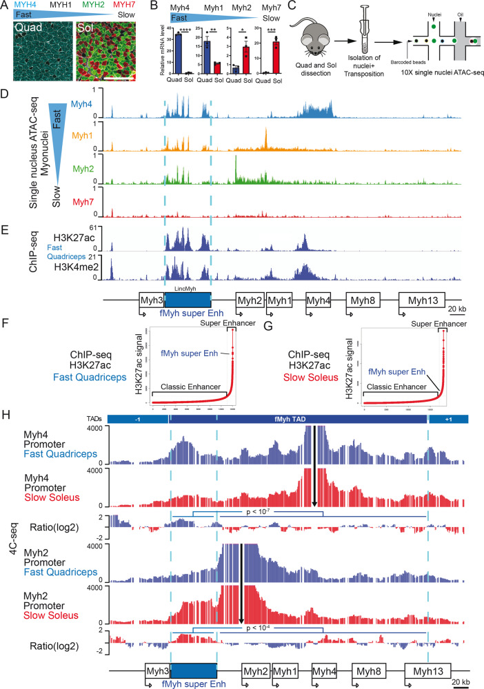
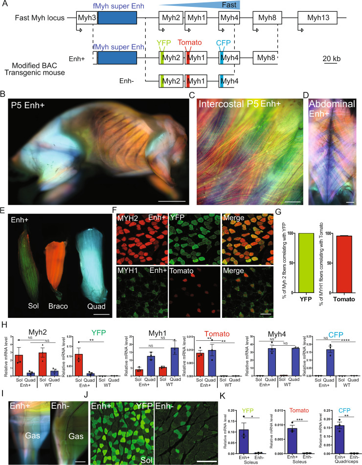
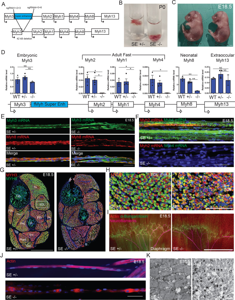
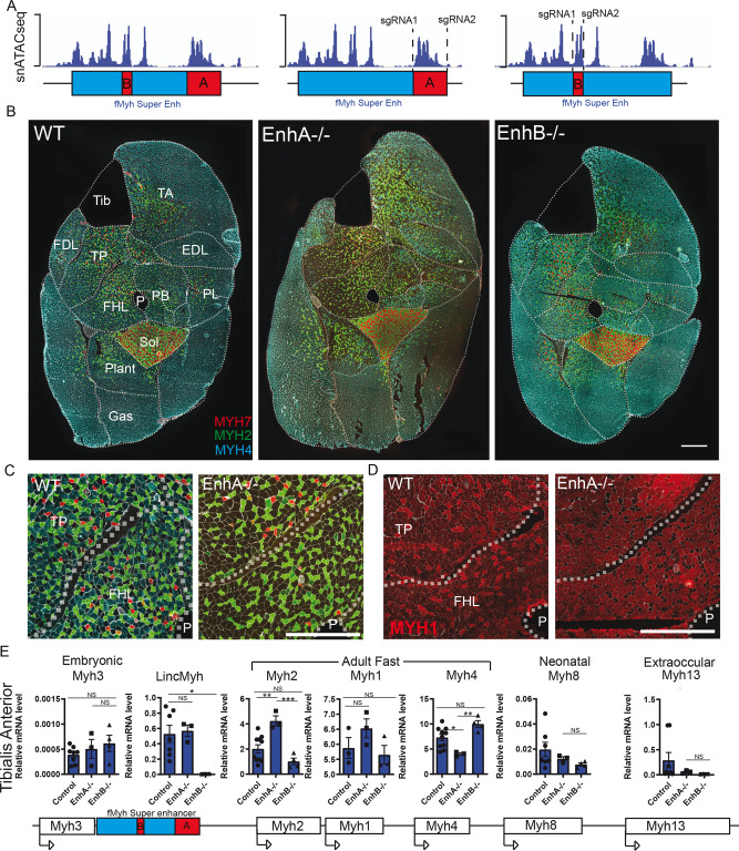
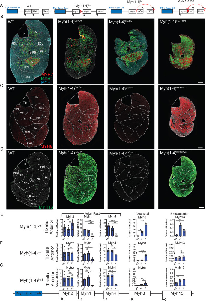
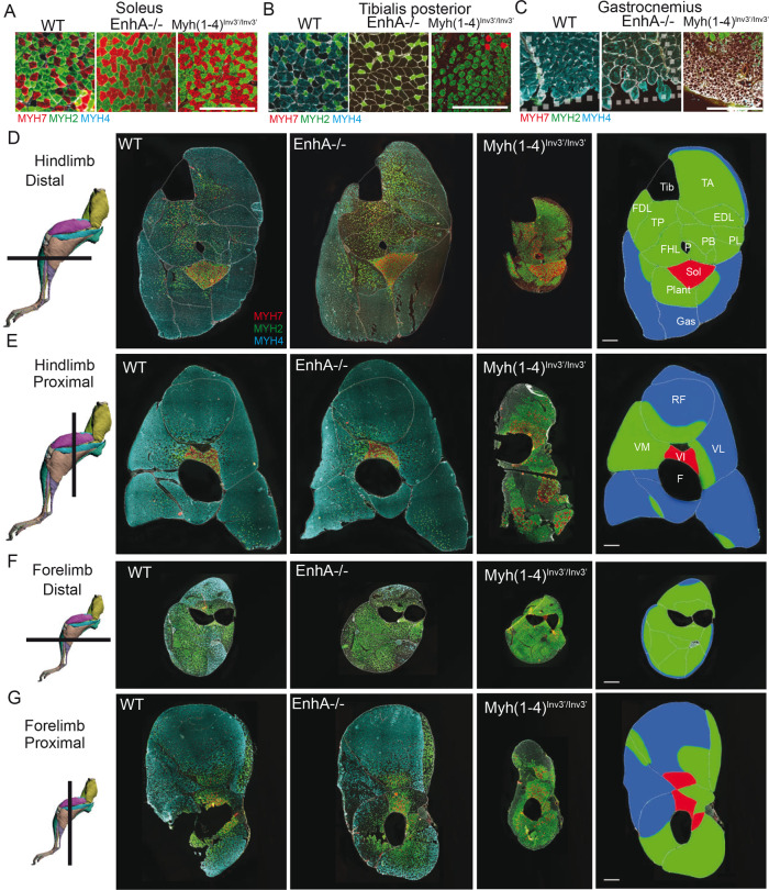
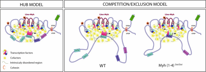
The contractile properties of adult myofibers are shaped by their Myosin heavy chain isoform content. Here, we identify by snATAC-seq a 42 kb super-enhancer at the locus regrouping the fast Myosin genes. By 4C-seq we show that active fast Myosin promoters interact with this super-enhancer by DNA looping, leading to the activation of a single promoter per nucleus. A rainbow mouse transgenic model of the locus including the super-enhancer recapitulates the endogenous spatio-temporal expression of adult fast Myosin genes. In situ deletion of the super-enhancer by CRISPR/Cas9 editing demonstrates its major role in the control of associated fast Myosin genes, and deletion of two fast Myosin genes at the locus reveals an active competition of the promoters for the shared super-enhancer. Last, by disrupting the organization of fast Myosin, we uncover positional heterogeneity within limb skeletal muscles that may underlie selective muscle susceptibility to damage in certain myopathies.


Conflict of interest statement

The authors declare no competing interests.

Figures

Fig. 1
Fig. 1. Identification of a super enhancer in the intergenic region of Myh3 and Myh2.
A Adult myofibers express different MYH isoforms. Immunostaining against fast (MYH4, MYH2) and slow (MYH7) MYH of adult fast quadriceps (Quad) and slow soleus (Sol) muscle sections, MYH1 + myofibers by default appear black. B Quantification by RT-qPCR of Myh mRNA expression in adult Quad and Sol (n = 3). C Graphical scheme of the experiments used for snATAC-seq experiments performed with slow soleus and fast quadriceps adult skeletal muscle. D Chromatin accessibility of the different types of myonuclei in the fast Myh locus. In fast myonuclei, we identified a 42-kb region with multiple chromatin accessibility peaks in the intergenic region of Myh3 and Myh2 genes. In slow Myh7 myonuclei this region of chromatin is not accessible. E H3K27Ac and H3K4me2 ChIP-seq signals were highly enriched in the 42 kb region of snATAC-seq peaks in the intergenic region of Myh3 and Myh2 genes. F Distribution of H3K27ac ChIP-seq signals across quadriceps and soleus enhancers. SEs contain high amounts of H3K27ac and the fMyh 42 kb sequence is identified as a SE. G Same as F in soleus. H 4C-seq experiments showing the interactions of the Myh4 (up) and Myh2 (down) promoters in quadriceps (blue) and soleus (red). Viewpoints are indicated by black arrows. The Ratio of interactions between the quadriceps and the soleus is indicated in between and shows that promoters of the active gene at the locus display significantly more interactions within the 42 kb cis-regulatory fMyh super enhancer. Significance of difference: G-test. For B, significance of difference by Student t test. Numerical data are presented as mean ± s.e.m. *P < 0.05, **P < 0.01, ***P < 0.001, ****P < 0.0001. Scale bars: 100 μm for A. Source data are provided as a Source Data file.
Fig. 2
Fig. 2. Transgenic models to study fMyh genes expression.
A Schematic representation of mouse fMyh locus and the recombined 222 kb Bacterial Artificial Chromosome (BAC) of the same locus. YFP, Tomato, and CFP cDNAs were inserted in the first exon of Myh1, Myh2, and Myh4 genes respectively in the BAC. Two transgenic mouse lines were obtained, one called Enh + that integrated 2 complete copies of the BAC and the other called Enh- devoid of the SE region and the 3′ region of the locus. The transgenes YFP, Tomato, and CFP are not to scale. BD Pictures of Enh + transgenic mice, red; Tomato, green; YFP and blue; CFP. All skeletal muscles expressed the transgenes. B 5-day-old lateral view. C zoom in intercostal muscles. D 2-month-old intercostal and abdominal muscles. E Transgene expression in adult soleus (Sol), bracoradial (Braco), and quadriceps (Quad) showing predominant expression of YFP in green, Tomato in red, and CFP in blue for each muscle. F Expression of the transgenes correlates with endogenous MYH protein expression in Enh+ line. Up: immunofluorescence against endogenous MYH2 (red) and of YFP (green) in adult soleus transverse section of Enh + mice. Down: immunofluorescence against endogenous MYH1 (green) and of Tomato (red) in adult quadriceps transverse section of Enh + mice. G Quantification of the percentage of MYH2 or MYH1 fibers expressing YFP or Tomato respectively, (n = 3). All MYH2 fibers are YFP + and almost all MYH1 fibers are Tomato+. H Relative expression level of mRNA in adult Sol and Quad of endogenous Myh genes and of transgenes, in wild type (WT) and in Enh + mice (n = 3). I Pictures of the adult leg of Enh + (left) and Enh- (right) mouse. The expression of the three transgenes is much higher in the Enh + line compared to Enh- mouse. J Immunostaining with GFP antibodies revealing YFP fibers on a section of adult Sol in Enh+ and Enh- mice. In Enh+ mouse, all MYH2 fibers expressed YFP whereas in Enh- only 10% of MYH2 fibers expressed YFP. K Quantification by RT-qPCR of transgenes expression in Enh+ and Enh− mouse line. Numerical data are presented as mean ± S.E.M. *P < 0.05, **P < 0.01, ***P < 0.001. Significance of difference, for H: two-way ANOVA and Student’s t test for K. Scale bars: 100 μm for F, and 50 μm for J. Source data are provided as a Source Data file.
Fig. 3
Fig. 3. The fMyh-SE is required for adult fMyh and neonatal Myh8 genes expression.
A A mouse line deleted for the fMyh-SE element was generated by injecting specific sgRNAs and Cas9 protein into mouse oocytes. B fMyh-SE−/− mice died at birth (P0) without breathing and air in their lungs. C fMyh-SE−/− E18.5 fetuses did not present severe visible malformations. D Quantification of Myh mRNAs by RT-qPCR in control and fMyh-SE−/− E18.5 forelimb skeletal muscles. Mutant muscles showed decreased Myh2, Myh1, Myh4, and Myh8 mRNAs levels. E RNAscope experiments against Myh3 and Myh8 mRNAs on isolated E18.5 forelimb fibers of control and mutant mice. F Same as E showing a decreased accumulation of Myh2 and Myh4 mRNAs in mutant mice compared to their littermate controls. G, H Immunostaining at the distal hindlimb level of E18.5 control and mutant fetuses revealing MYH3 and MYH8 positive myofibers. H zoom in the EDL of control and mutant fetuses. I In toto immunostaining of diaphragms from E18.5 mutant and control fetuses showing in red Actin filaments (phalloidine), in green AchR (alpha-bungarotoxin), and in pink neurofilaments. Mutant diaphragms show altered repartition of NMJ and punctated Actin aggregates. J Myofibers from mutant diaphragm showed defects in sarcomeres organization as shown by phalloidine staining. K Electronic microscopy pictures of the sarcomeres defects present in mutant E18.5 fetuses compared to their littermate controls. For D (n = 3). For E and F, scale bar: 50 μm. For G, scale bar: 500 μm. For H, scale bar: 100 μm, 500 μm for I and 25 μm for K. Numerical data are presented as mean ± s.e.m. *P < 0.05, **P < 0.01. Significance of difference, for D: one-way ANOVA with multiple comparisons. Source data are provided as a Source Data file.
Fig. 4
Fig. 4. Role of the different enhancers composing the SE.
A Schematic representation of the snATAC-seq peaks along the 42 kb SE and the enhancers A and B deleted by CRISPR/Cas9 editing. B Immunostaining against fast MYH2, MYH4, and slow MYH7 on adult leg sections of 2–3-month-old mouse female deleted for enhancer A or B. C Same as B, zoom in Tibialis posterior and FHL muscle of WT and EnhA−/− mutant. D Immunostaining against fast MYH1 in Tibialis posterior and FHL muscle of WT and EnhA−/− mutant. The absence of EnhA induced an increased number of MYH1 positive fibers. E Quantification of fMyh mRNA and of Linc-Myh in adult TA of control and EnhA and EnhB mutant by RT-qPCR experiments. For E, n = 3. Numerical data are presented as mean ± S.E.M. *P < 0.05, **P < 0.01, ***P < 0.001. Scale bars: 100 μm for BD. Significance of difference, for E: one-way ANOVA with multiple comparisons. Source data are provided as a Source Data file.
Fig. 5
Fig. 5. The promoters of fMyh genes compete for the shared SE.
A Schema of the distinct fMyh alleles generated by CRISPR/Cas9 editing. B Immunostaining against MYH4 (blue), MYH2 (green), and slow MYH7 (red) of adult distal hindlimb sections of WT, Myh(1–4)Del/Del, Myh(1–4)Inv/Inv, and of Myh(1–4)Inv3Inv3 mutants. C Immunostaining against neonatal MYH8 of adult leg sections in WT, Myh(1–4)Del/Del, Myh(1–4)Inv/Inv, and of Myh(1–4)Inv3′/Inv3. D Same as C against extraocular MYH13. E Quantification of Myh2, Myh1, Myh4, Myh8, and Myh13 mRNAs of adult WT, Myh(1–4)Del/+ and Myh(1–4)Del/Del TA by RT-qPCR experiments. F Quantification of Myh2, Myh1, Myh4, Myh8, and Myh13 mRNAs of adult WT, Myh(1–4)Inv/+ and Myh(1–4)Inv/Inv TA by RT-qPCR experiments. G Quantification of Myh2, Myh1, Myh4, Myh8, and Myh13 mRNAs of adult WT, Myh(1–4)Inv3/+ and Myh(1–4)Inv3′/Inv3 TA by RT-qPCR. For EG (n = 3). Numerical data are presented as mean ± S.E.M. *P < 0.05, **P < 0.01, ***P < 0.001. Significance of difference, for CE: one-way ANOVA with multiple comparisons. Scale bars: 50 μm for G. Source data are provided as a Source Data file.
Fig. 6
Fig. 6. MYH expression established different groups of limb skeletal muscles in mutant animals.
AC Immunostaining against MYH7 (red), MYH2 (green), and MYH4 (blue) in WT, EnhA−/−, and Myh(1–4)Inv3′/Inv3adult mice. The soleus (A) is not affected in these mutant mice. In the Tibialis posterior (B), the expression of MYH4 is lost in EnhA−/−, and in Myh(1–4)inv3’/inv3’ whereas an upregulation of MYH1 is observed in EnhA−/− mice and an upregulation of MYH2 in Myh(1–4)Inv3′/Inv3 mice. In Gastrocnemius (C), the peripheral fibers of Myh(1–4)Inv3′/Inv3 mice present a severe atrophy. In contrast these fibers are not affected in EnhA−/− mouse. D Comparison of the phenotypes of adult muscles in EnhA−/− and in Myh(1–4) Inv3′/Inv3allowed classification of distal hindlimb muscles in three major categories. The first group is shown in red corresponded to the soleus that is not affected. The second group shown in green corresponded to muscles affected in EnhA−/− and Myh(1–4)Inv3′/Inv3 mutants. The third group shown in blue corresponded to muscles strongly affected in Myh(1–4)Inv3′/Inv3 but not in EnhA−/− mutants. E Same as D in the proximal part of the hindlimb. F Same as D in the distal part of the forelimb. G Same as D in the proximal part of the forelimb. For D, scale bar: 100 μm. DG: drawings of hindlimbs and forelimbs are from Charles et al..
Fig. 7
Fig. 7. Two models explaining the complex regulation of fMyh genes by the shared SE.
The SE is composed of seven enhancer elements (1–7) recruiting TF and cofactors allowing the nuclear formation of a phase separation condensate in myonuclei and allowing robust fMyh expression, adapted from Sabari et al.. Left, in the hub model even the inactive promoters at the locus are associated in the phase-separated droplet in Myh4 + myonuclei. Right, in the competition/exclusion model, Myh2, Myh1, Myh8, and Myh13 are excluded from the phase-separated droplet in Myh4 + myonuclei because they are not bound by sufficient amount of TF and cofactors. In those myonuclei robust bi-allelic expression of Myh4 is achieved, while transcription of the other genes is not detected. In this competition/exclusion model, the deletion of Myh1 and Myh4 could lead to the maintenance of Myh8 expression through development in specific muscles, (while in others Myh13 is activated (not represented)) due to the absence of competition by Myh4 or Myh1 promoters. Linc-Myh spans enhancers 3–5. The models are not to scale.

Similar articles

Cited by

References

    1. Emery AE. Muscular dystrophy into the new millennium. Neuromuscul. Disord. 2002;12:343–349. - PubMed
    1. Mantilla CB, Sieck GC. Invited review: mechanisms underlying motor unit plasticity in the respiratory system. J. Appl. Physiol. 2003;94:1230–1241. - PubMed
    1. Kanning KC, Kaplan A, Henderson CE. Motor neuron diversity in development and disease. Annu. Rev. Neurosci. 2010;33:409–440. - PubMed
    1. Schiaffino S, Reggiani C. Fiber types in mammalian skeletal muscles. Physiol. Rev. 2011;91:1447–1531. - PubMed
    1. Gundersen K. Excitation-transcription coupling in skeletal muscle: the molecular pathways of exercise. Biol. Rev. Camb. Philos. Soc. 2011;86:564–600. - PMC - PubMed

Publication types