. 2022 Feb 24;5(1):166.
doi: 10.1038/s42003-022-03110-8.

An amiloride derivative is active against the F1Fo-ATP synthase and cytochrome bd oxidase of Mycobacterium tuberculosis

Affiliations

An amiloride derivative is active against the F1Fo-ATP synthase and cytochrome bd oxidase of Mycobacterium tuberculosis

Kiel Hards et al. Commun Biol. .

Abstract

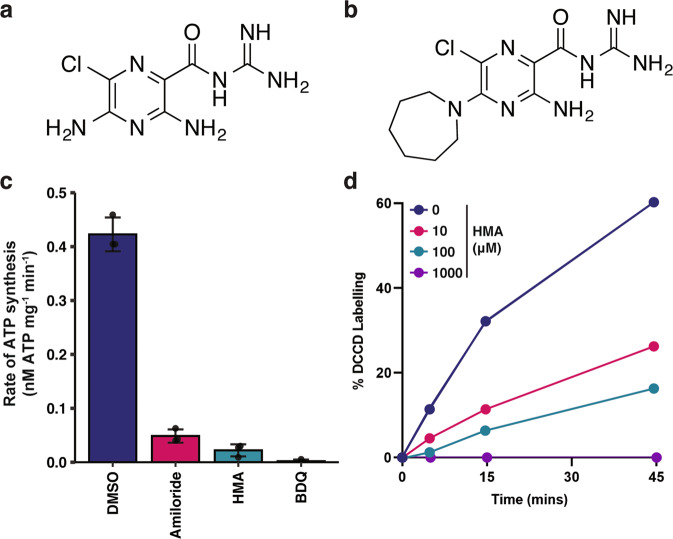
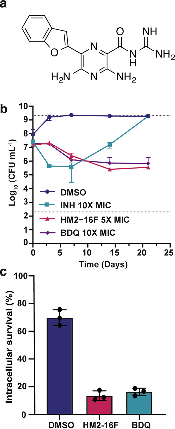
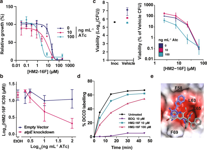
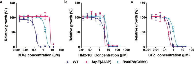
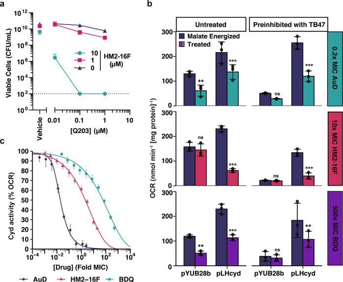
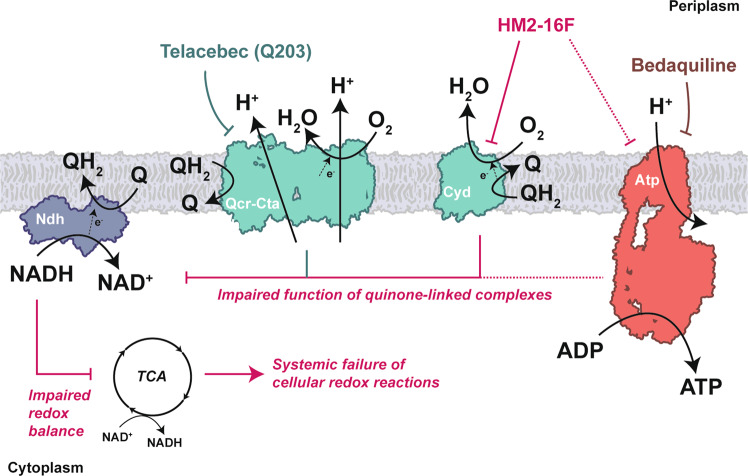
Increasing antimicrobial resistance compels the search for next-generation inhibitors with differing or multiple molecular targets. In this regard, energy conservation in Mycobacterium tuberculosis has been clinically validated as a promising new drug target for combatting drug-resistant strains of M. tuberculosis. Here, we show that HM2-16F, a 6-substituted derivative of the FDA-approved drug amiloride, is an anti-tubercular inhibitor with bactericidal properties comparable to the FDA-approved drug bedaquiline (BDQ; Sirturo®) and inhibits the growth of bedaquiline-resistant mutants. We show that HM2-16F weakly inhibits the F1Fo-ATP synthase, depletes ATP, and affects the entry of acetyl-CoA into the Krebs cycle. HM2-16F synergizes with the cytochrome bcc-aa3 oxidase inhibitor Q203 (Telacebec) and co-administration with Q203 sterilizes in vitro cultures in 14 days. Synergy with Q203 occurs via direct inhibition of the cytochrome bd oxidase by HM2-16F. This study shows that amiloride derivatives represent a promising discovery platform for targeting energy generation in drug-resistant tuberculosis.


Conflict of interest statement

The authors declare no competing interests.

Figures

Fig. 1
Fig. 1. Inhibition of the mycobacterial F1Fo-ATP synthase by amiloride and hexamethylene amiloride.
a Structure of amiloride and b 5-(N,N-hexamethylene)amiloride (HMA). c ATP synthesis activity of NADH-energized M. smegmatis inverted membrane vesicles (IMV), treated with either 64 μM of amiloride, 64 μM of HMA, or 2 μM of BDQ as indicated. Error bars represent standard deviation from triplicate biologically independent measurements. d Time course of DCCD labelling in purified M. phlei c-rings in the presence of the indicated amounts of HMA. Data are representative of duplicate measurements.
Fig. 2
Fig. 2. HM2-16F is bactericidal towards mycobacteria in vitro and in macrophages.
a Structure of HM2-16F. b Survival of M. tuberculosis after treatment with: isoniazid (INH 2 μM, 10× MIC), HM2-16F (20 μM, 5× MIC) and bedaquiline (BDQ 2 μM, 10X MIC). c Survival of M. bovis BCG in human THP-1 macrophages after treatment with 5 μM HM2-16F and BDQ. Error bars indicate standard deviation from three biological independent experiments.
Fig. 3
Fig. 3. Interactions of HM2-16F with the mycobacterial F1Fo-ATP synthase.
a Knockdown of the ATP synthase operon (sgRNA targeting atpB – pCi74) was induced with the indicated amounts of ATc and the growth relative to the vehicle control was determined. Error bars represent standard deviation from three biological independent experiments. b Growth after 10 days and IC50 of HM2-16F as a function of F1Fo-ATP synthase knockdown (sgRNA targeting atpB – pCi74). Error bars represent 95% confidence interval. c Viability of M. tuberculosis after 10 days was determined in an analogous experiment to panel a at 0, 10 and 100 ng mL1. d Time course of DCCD labelling in purified M. phlei c-rings in the presence of the indicated amounts of compounds. Data are representative of duplicate measurements. e HM2-16F (purple carbons) docked into the BDQ binding site of mycobacterial F1Fo-ATP synthase c-ring (PDB ID: 4V1F). The high-resolution c-ring-BDQ structure is shown in grey as a reference. The protein is shown as an electrostatic potential surface (red – electronegative, white – neutral, blue – electropositive; generated in PyMOL). Amino acid positions refer to the M. phlei protein. The top three ranked poses are shown in Supplementary Figure 5.
Fig. 4
Fig. 4. Bedaquiline-resistant mutants of M. tuberculosis are not cross-resistant to HM2-16F.
The M. tuberculosis strains mc26206 (WT), AtpE(A63P), and Rv0678(G65fs) were grown in the presence of increasing concentrations of: a BDQ, b HM2-16F or c clofazimine and growth measured after 10 days. Error bars represent standard deviation from four biological independent experiments.
Fig. 5
Fig. 5. Metabolomic profiling of HM2-16F-treated cultures of M. tuberculosis indicates HM2-16F inhibits respiration and induces cellular reductive stress.
a Global profile of metabolites with dose-dependent changes, as assessed by log-linear regressions. b Global profile of metabolites with dose-independent changes, as assessed by two sample t-tests with Benjamini & Hochberg False Discovery Rate (FDR) adjustment. c Fold changes of selected metabolites, classified according to whether the metabolite changed in a dose-dependent (increasing/decreasing) or dose-independent manner. d Changes in adenine nucleotide/nucleoside pools. e Relative changes in the indicated metabolites. Raw data are available in Supplementary Data 1.
Fig. 6
Fig. 6. HM2-16F synergizes with Q203 by inhibiting cytochrome bd oxidase.
a Cell killing (viability) of M. tuberculosis with increasing amounts of both Q203 and HM2-16F after 14 days challenge with both compounds. Error bars indicate standard deviation from three independent experiments. b Inhibition of the M. tuberculosis cytochrome bd oxidase in IMVs from M. smegmatis mc2155 ΔcydAB with either pYUB28b (empty vector) or pLHcyd- MtbCydABDC+. IMVs were pre-inhibited with 500 nM TB47 for 1 min as indicated. Oxygen consumption was initiated with 5 mM malate (final concentration) as the sole electron donor. The indicated treatment: 0.2× MIC aurachin D (AuD) (top panel 6b), 10× MIC HM2-16F (middle panel b) and 500× MIC BDQ (bottom panel b) was added once malate energized oxygen consumption reached steady state (~2 min after malate addition). Error bars represent standard deviation from triplicate measurements. Asterisks indicate significance compared to the corresponding malate energized OCR *** = p < 0.001, ** = p < 0.01, ns = p > 0.05 (One-way ANOVA, Tukey multiple comparisons, 95% CI). c Titration of the indicated compounds in malate-energized IMVs of M. smegmatis mc2155 ΔcydAB pLHcyd- MtbCydABDC+ pre-inhibited with 500 nM TB47 (final concentration). Error bars indicate standard error from four technical replicates.
Fig. 7
Fig. 7. Summary of the action of HM2-16F at the level of the respiratory chain.
HM2-16F inhibits cytochrome bd and, to a lesser or indirect extent, the F1Fo-ATP synthase. Disruption of quinol regeneration can affect the primary dehydrogenases of the respiratory chain leading to dysregulated regeneration of reducing equivalents that can lead to a systemic failure of cellular redox reactions. The addition of Q203 (or TB47), direct inhibitors of the cytochrome bcc-aa3 oxidase branch, removes the only alternative pathway that mycobacteria can use to escape this inhibition of oxygen consumption leading to cell death.

References

    1. World Health Organization. Global Tuberculosis Report 2019 (Geneva: World Health Organization, 2019).
    1. Dheda K, et al. Global control of tuberculosis: from extensively drug-resistant to untreatable tuberculosis. Lancet Respir. Med. 2014;2:321–338. - PMC - PubMed
    1. World Health Organization. Guidelines for the Treatment of Tuberculosis: Fourth Edition (World Health Organization, 2010).
    1. World Health Organization. Guidelines for the Programmatic Management of Drug-Resistant Tuberculosis: Emergency Update 2008 (Geneva: World Health Organization, 2008).
    1. Jones D. Tuberculosis success. Nat. Rev. Drug Discov. 2013;12:175–176. - PubMed

MeSH terms