. 2022 Feb 24;5(1):165.
doi: 10.1038/s42003-022-03074-9.

Molecular dynamics simulations suggest possible activation and deactivation pathways in the hERG channel

Affiliations

Molecular dynamics simulations suggest possible activation and deactivation pathways in the hERG channel

Flavio Costa et al. Commun Biol. .

Abstract

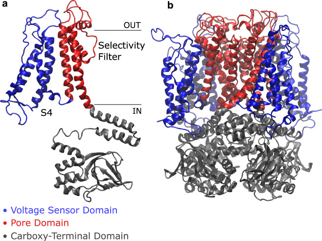
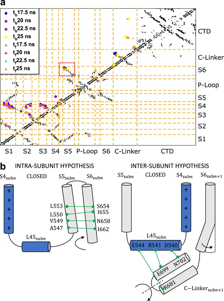
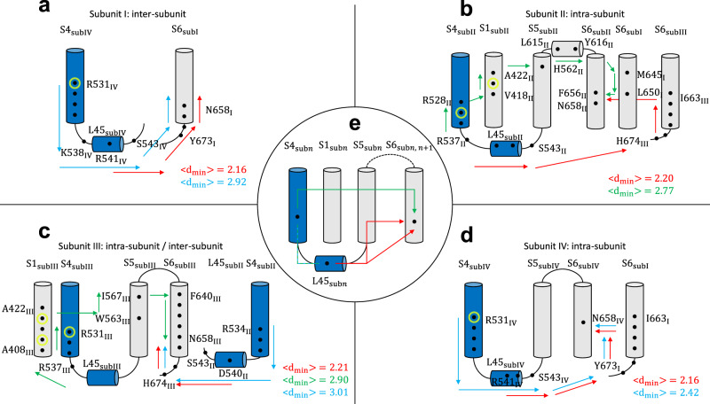
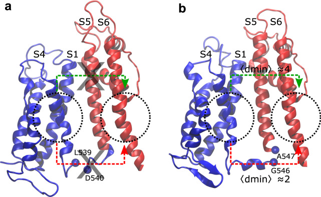
The elusive activation/deactivation mechanism of hERG is investigated, a voltage-gated potassium channel involved in severe inherited and drug-induced cardiac channelopathies, including the Long QT Syndrome. Firstly, the available structural data are integrated by providing a homology model for the closed state of the channel. Secondly, molecular dynamics combined with a network analysis revealed two distinct pathways coupling the voltage sensor domain with the pore domain. Interestingly, some LQTS-related mutations known to impair the activation/deactivation mechanism are distributed along the identified pathways, which thus suggests a microscopic interpretation of their role. Split channels simulations clarify a surprising feature of this channel, which is still able to gate when a cut is introduced between the voltage sensor domain and the neighboring helix S5. In summary, the presented results suggest possible activation/deactivation mechanisms of non-domain-swapped potassium channels that may aid in biomedical applications.


Conflict of interest statement

The authors declare no competing interests.

Figures

Fig. 1
Fig. 1. Side view of hERG channel.
A single subunit (a) and the whole protein (b) are colored by domain: in blue, the voltage sensor domain ranging from helix S1 to loop L45, including the positively charged helix S4; in red, the pore domain, composed of helix S5, P-Loop, selectivity filter, and helix S6; in gray, the carboxy-terminal domain with the C-Linker and the cyclic nucleotide-binding homology domain.
Fig. 2
Fig. 2. Contact analysis and activation/deactivation hypotheses.
a Contact map of the first subunit of the system with Qg = 8e during the transition from O to C state. The black dots represent the contacts in the initial O conformation not broken during the simulation while the colored symbols indicate the contacts formed or broken at different times (tf and tb, respectively) during TMD. b Illustration of the two activation/deactivation hypotheses displaying the relevant conserved interactions in the C state.
Fig. 3
Fig. 3. Activation/deactivation pathways of the closed system with Qg = 8e.
Paths identified in subunits I (a), II (b), III (c), and IV (d). Panel e schematizes the main families of activation/deactivation pathways identified in all systems. Arrows describe the preferred routes of motion propagation: blue arrows refer to S4→L45→S6 route; red arrows refer to the L45→S6 route; green arrows refer to S4→S1→S5→S6. Black dots correspond to residues on the path with CI > 0.15 (see Supplementary Table 1); yellow circles refer to the pathological mutation R531Q/W known to alter the gating of the channel inducing the LQTS. The mutations W410S, Y420C, and T421M impair both the trafficking and the gating. Average minimal path lengths dmin are also reported.
Fig. 4
Fig. 4. Communication pathways in split channels.
Split channels disconnected before D540 (a) and after G546 (b): dotted circles are centered on the source and sink regions used in the network analysis; these locations correspond to helices S4 and S6; red arrows schematize the S4→L45→S6 and S4→L45→S5→S6 paths while green arrows refer to S4→S1→S5→S6 path. Average minimal path lengths dmin are reported in the G546-split system (b); no pathways are found for D540 (a).
Fig. 5
Fig. 5. Pseudo-potential of mean force for hERG with Qg = 8e as a function of geometrical descriptors, relevant for the O→C transition.
The geometrical observables are: displacement and rotation of helices S4 and bending of helices S6 (a); rotation of the cytosolic domains with respect to the transmembrane ones (b). Pseudo-PMF maps as follows: S4 rotation vs S4 displacement (c); S6 bending vs S4 displacement (d); S6 bending vs S4 rotation (e); PD-CTD rotation vs S6 bending (f); PD-CTD rotation vs S4 displacement (g); PD-CTD rotation vs S4 rotation (h). Dashed lines are a guide to the eye. Bin widths are as follows: S4 displacement: 1.0 Å; S4 rotation: 2°; S6 bending: 1°; PD-CTD rot: 1°. Free energies were expressed in kcal/mol.

Similar articles

Cited by

References

    1. Trudeau MC, Warmke JW, Ganetzky B, Robertson GA. HERG, a human inward rectifier in the voltage-gated potassium channel family. Science. 1995;269:92–95. - PubMed
    1. Curran ME, et al. A molecular basis for cardiac arrhythmia: hERG mutations cause long QT syndrome. Cell. 1995;80:795–803. - PubMed
    1. Sanguinetti MC, Tristani-Firouzi M. hERG potassium channels and cardiac arrhythmia. Nature. 2006;440:463–469. - PubMed
    1. Recanatini M, Poluzzi E, Masetti M, Cavalli A, De Ponti F. QT prolongation through hERG K+ channel blockade: current knowledge and strategies for the early prediction during drug development. Med. Res. Rev. 2005;25:133–166. - PubMed
    1. Butler A, Helliwell MV, Zhang Y, Hancox JC, Dempsey CE. An update on the structure of hERG. Front. Pharmacol. 2020;10:1572. - PMC - PubMed

Publication types

MeSH terms

Substances