

Primary Grynfeltt Lumbar Hernia: A Case Report
- PMID: 35210629
- PMCID: PMC9199999
- DOI: 10.31729/jnma.7251
Primary Grynfeltt Lumbar Hernia: A Case Report
Abstract
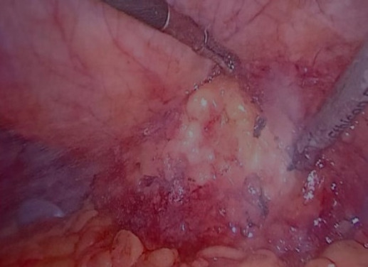
A weakening or defect in posterolateral abdominal wall can lead to development of lumbar hernia. These defects are particularly common in Petit's inferior triangle or Grynfeltt-Lesshaft superior triangle. There are very few cases of primary lumbar hernias that have been described in literature till date. As it is a rare entity, it is often misdiagnosed, leading to delay in management. We present a case of a 66-year-old male with no previous surgery who presented with a mass in left lumbar region for last ten years. The mass gradually increased in size and caused vague dragging pain. On Computed tomography, the diagnosis of Grynfeltt hernia was made. The patient underwent a laparoscopic mesh repair and had an uneventful postoperative hospital stay. Although a rare entity, there should be a high degree of suspicion of a lumbar hernia when evaluating a case of a lumbar mass. Early diagnosis by computed tomography and management with open or minimally invasive techniques can prevent complications.
Keywords: abdominal hernia; case report; surgical mesh..
Conflict of interest statement
Figures


Similar articles
-
Laparoscopic repair of a lumbar hernia: report of a case and extensive review of the literature.Surg Endosc. 2013 Sep;27(9):3421-9. doi: 10.1007/s00464-013-2884-9. Epub 2013 Apr 30. Surg Endosc. 2013. PMID: 23636518
-
Grynfelt hernia: case report and literature review.Hernia. 2012 Feb;16(1):107-11. doi: 10.1007/s10029-010-0722-8. Epub 2010 Sep 5. Hernia. 2012. PMID: 20821030 Review.
-
Robotic-Assisted Laparoscopic Repair of Petit's Hernia With Preperitoneal Mesh.Cureus. 2024 Jul 3;16(7):e63771. doi: 10.7759/cureus.63771. eCollection 2024 Jul. Cureus. 2024. PMID: 38966780 Free PMC article.
-
Lumbar hernia: surgical anatomy, embryology, and technique of repair.Am Surg. 2009 Mar;75(3):202-7. Am Surg. 2009. PMID: 19350853 Review.
-
Primary lumbar hernia repair: the open approach.Eur Surg Res. 2007;39(2):88-92. doi: 10.1159/000099155. Epub 2007 Feb 1. Eur Surg Res. 2007. PMID: 17283432 Clinical Trial.
Cited by
-
Impact of Refractive Status on Presbyopia Progression among Patients with Presbyopia.Graefes Arch Clin Exp Ophthalmol. 2024 Aug;262(8):2695-2701. doi: 10.1007/s00417-024-06455-4. Epub 2024 Mar 21. Graefes Arch Clin Exp Ophthalmol. 2024. PMID: 38512509
-
A novel mini-open sublay hernioplasty combined with D10 mesh for primary lumbar hernia: a retrospective analysis of 48 cases.Hernia. 2023 Oct;27(5):1283-1288. doi: 10.1007/s10029-023-02812-0. Epub 2023 Jun 6. Hernia. 2023. PMID: 37277523
References
-
- Klingensmith ME, Wise PE. The Washington manual of surgery. 8th ed. Alphen on the Rhine: Wolters Kluwer; 2020. Chapter 29, Hernias.https://books.google.com.np/books?id=PN3zugEACAAJ&dq=%22The+Washington+M... Available from:
Publication types
MeSH terms
LinkOut - more resources
Full Text Sources
Research Materials