. 2022 Jan-Feb;55(1):47-53.
doi: 10.1590/0100-3984.2020.0141.

The 2017 ACR TI-RADS: pictorial essay

Affiliations

The 2017 ACR TI-RADS: pictorial essay

André Tojal Pires et al. Radiol Bras. 2022 Jan-Feb.

Abstract

High-resolution ultrasound is the imaging method of choice for the evaluation of thyroid nodules. The method has recently come to be used widely and often, which has increased the rate of thyroid nodule detection. In 2017, the American College of Radiology (ACR) established a risk-stratification system designated the Thyroid Imaging Reporting and Data System (TI-RADS) to be a practical guide for widespread use, with a single lexicon and standardization of ultrasound reports of thyroid nodules. The objective of this study was to present a practical approach, using examples to illustrate the criteria evaluated by the 2017 ACR TI-RADS, in order to help minimize uncertainties regarding its application by sonographers.

A ultrassonografia de alta resolução é a modalidade de escolha para avaliação de imagem dos nódulos tireoidianos, e sua recente aplicação ampla e difusa tornou a detecção de nódulos tireoidianos mais frequentes. O American College of Radiology (ACR) estabeleceu um sistema de estratificação de risco denominado Thyroid Imaging, Reporting and Data System (TI-RADS) para ser um guia prático para utilização ampla com um léxico único e padronização de relatórios ultrassonográficos de nódulos tireoidianos. O objetivo deste trabalho é fazer uma abordagem prática com base em exames para ilustrar e exemplificar os critérios avaliados pelo TI-RADS-ACR 2017, a fim de ajudar a reduzir os pontos de dúvidas de sua aplicação pelos profissionais ultrassonografistas.

Keywords: Thyroid diseases; Thyroid gland; Ultrasonography.


Figures

Figure 1
Figure 1
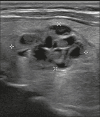
Image of a nodule that was completely cystic. Nodules that are completely cystic, predominantly cystic, or spongiform are not scored for other categories, therefore automatically receiving a final score of 0 and classified as TR1.
Figure 2
Figure 2
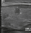
Image of a mixed solid-cystic nodule. In such nodules, only the solid component should be scored for the echogenicity, margin, and echogenic foci categories. In this case, the nodule was assigned 1 point for being mixed, 2 points for being hypoechoic, 0 points for being wider-than-tall, 0 points for having undefined margins, and 0 points for having no acoustic shadowing artifacts or echogenic foci. Therefore, the total score was 3 points and the risk level was classified as TR3.
Figure 3
Figure 3
Image of a completely solid nodule, with echogenicity similar to the rest of the thyroid parenchyma, presenting a hypoechoic halo that should not be scored for the echogenicity or margin categories. The features of (scores for) this nodule were as follows: solid (2 points); isoechoic (1 point); wider-than-tall (0 points); smooth margins (0 points); and no acoustic shadowing artifacts or echogenic foci (0 points). Therefore, the total score was 3 points and the risk level was classified as TR3.
Figure 4
Figure 4
Image of a hyperechoic nodule. The features of (scores for) this nodule were as follows: solid (2 points); hyperechoic (1 point); wider-than-tall (0 points); smooth margins (0 points); and no acoustic shadowing artifacts or echogenic foci (0 points). Therefore, the total score was 3 points and the risk level was classified as TR3.
Figure 5
Figure 5
Image of a hyperechoic nodule. Note the heterogeneous echotexture of the thyroid parenchyma, especially the presence of nodules with well-defined margins and echogenicity greater than that of the rest of the parenchyma. The nodule pictured was solid (2 points), hyperechoic (1 point), and wider-than-tall (0 points), with smooth margins (0 points) and without acoustic shadowing artifacts or echogenic foci (0 points). Therefore, the total score was 3 points and the risk level was classified as TR3.
Figure 6
Figure 6
Image of a predominantly solid nodule with smooth margins that is less echogenic than the rest of the thyroid parenchyma. The nodule pictured was solid (2 points), hypoechoic (2 points), and wider-than-tall (0 points), with well-defined margins (0 points) and without acoustic shadowing artifacts or echogenic foci (0 points). Therefore, the total score was 4 points and the risk level was classified as TR4.
Figure 7
Figure 7
Image of an oval-shaped nodule with well-defined margins and echogenicity lower than that of the rest of the thyroid parenchyma. In this case, the nodule was assigned 2 points for being solid, 2 points for being hypoechoic, 0 points for being wider-than-tall, 2 points for having a lobulated margin, and 0 points for having no acoustic shadowing artifacts or echogenic foci. Therefore, the total score was 6 points and the risk level was classified as TR4.
Figure 8
Figure 8
Image of a markedly hypoechoic nodule. Compare the echogenicity of the nodules with that of the cervical musculature. Attention should be paid to the ultrasound parameters. The nodule pictured was solid (2 points), markedly hypoechoic (3 points), and wider-than-tall (0 points), with undefined margins (0 points) and without acoustic shadowing artifacts or echogenic foci (0 points). Therefore, the total score was 5 points and the risk level was classified as TR4.
Figure 9
Figure 9
Image of a predominantly solid, hypoechoic, taller-than-wide nodule with well-defined margins. The features of (scores for) this nodule were as follows: solid (2 points), hypoechoic (2 points), taller-than-wide (3 points), smooth margins (0 points), and no acoustic shadowing artifacts or echogenic foci (0 points). Therefore, the total score was 7 points and the risk level was classified as TR5.
Figure 10
Figure 10
Image of a solid nodule with lobulated margins and a rounded lobulation in its anterior portion. In this case, the nodule was assigned 2 points for being solid, 2 points for being hypoechoic, 0 points for being wider-than-tall, 2 points for having a lobulated margin, and 0 points for having no acoustic shadowing artifacts or echogenic foci. Therefore, the total score was 6 points and the risk level was classified as TR4.
Figure 11
Figure 11
Image of a solid nodule with irregular margins. Note the irregularity, with an acute angle at the medial margin of the nodule. The nodule pictured was solid (2 points), hypoechoic (2 points), and wider-than-tall (0 points), with irregular margins (2 points) and without acoustic shadowing artifacts or echogenic foci (0 points). Therefore, the total score was 6 points and the risk level was classified as TR4.
Figure 12
Figure 12
Image of a nodule extending beyond the anterior limit of the thyroid. The nodule pictured was solid (2 points), hypoechoic (2 points), and wider-than-tall (0 points), with extrathyroidal extension (3 points) and without posterior attenuation artifacts or echogenic foci (0 points). Therefore, the total score was 7 points and the risk level was classified as TR5.
Figure 13
Figure 13
Image of a mixed solid-cystic nodule. Note that the medial margin of the nodule cannot easily be distinguished from the rest of the parenchyma. In this case, the nodule was assigned 1 point for being mixed, 2 points for being hypoechoic, 0 points for being wider-than-tall, 0 points for having ill-defined margins, and 0 points for having no acoustic shadowing artifacts or echogenic foci. Therefore, the total score was 3 points and the risk level was classified as TR3.
Figure 14
Figure 14
Image of a solid nodule, showing punctate echogenic foci. The nodule pictured was solid (2 points), hypoechoic (2 points), and wider-than-tall (0 points), with undefined margins (0 points) and punctate echogenic foci (3 points). Therefore, the total score was 7 points and the risk level was classified as TR5.
Figure 15
Figure 15
Image of a nodule with macrocalcification. Note the intense acoustic shadowing. The features of (scores for) this nodule were as follows: solid (2 points); isoechoic (1 point); wider-than-tall (0 points); smooth margins (0 points); and macrocalcification (1 point). Therefore, the total score was 4 points and the risk level was classified as TR4.
Figure 16
Figure 16
Image of a nodule with peripheral echogenic foci corresponding to calcifications. The nodule pictured was solid (2 points), hypoechoic (2 points), and wider-than-tall (0 points), with well-defined margins (0 points) and peripheral calcifications (2 points). Therefore, the total score was 6 points and the risk level was classified as TR4.
Figure 17
Figure 17
Image of a nodule with peripheral calcifications and acoustic shadowing that obscures its central content. According to the ACR TI-RADS, when the internal characteristics of a nodule cannot be determined because of acoustic shadowing, it is prudent to assume that it is solid and to assign it 2 points for composition, as well as 1 point for echogenicity. In this case, the nodule was assigned 2 points for being of indeterminate composition, 1 point for being of indeterminate echogenicity, 0 points for being wider-than-tall, 2 points for having lobulated margins, and 2 points for having peripheral calcifications. Therefore, the total score was 7 points and the risk level was classified as TR5.
Figure 18
Figure 18
Image of a nodule that was solid (2 points), isoechoic (1 point), and wider-than-tall (0 points), with smooth margins (0 points) and without echogenic foci or acoustic shadowing artifacts (0 points). Therefore, the total score was 3 points and the risk level was classified as TR3.
Figure 19
Figure 19
Image of a nodule that was solid (2 points), markedly hypoechoic (3 points), and wider-than-tall (0 points), with smooth margins (0 points) and without echogenic foci or acoustic shadowing artifacts (0 points). Therefore, the total score was 5 points and the risk level was classified as TR4.
Figure 20
Figure 20
Image of a mixed solid-cystic nodule (1 point) that was isoechoic (1 point), was wider-than-tall (0 points), and extended beyond the anterior limit of the thyroid gland (3 points), without echogenic foci or acoustic shadowing artifacts (0 points). Therefore, the total score was 5 points and the risk level was classified as TR4.
Figure 21
Figure 21
Image of a nodule that was solid (2 points), hypoechoic (2 points), and taller-than-wide (3 points), with undefined margins (0 points) and without acoustic shadowing artifacts or echogenic foci (0 points). Therefore, the total score was 7 points and the risk level was classified as TR5.

Similar articles

Cited by

References

    1. Ezzat S, Sarti DA, Cain DR, et al. Thyroid incidentalomas. Prevalence by palpation and ultrasonography. Arch Intern Med. 1994;154:1838–1840. - PubMed
    1. Nam-Goong IS, Kim HY, Gong G, et al. Ultrasonography-guided fine-needle aspiration of thyroid incidentaloma: correlation with pathological findings. Clin Endocrinol (Oxf) 2004;60:21–28. - PubMed
    1. Ito Y, Miyauchi A, Inoue H, et al. An observational trial for papillary thyroid microcarcinoma in Japanese patients. World J Surg. 2010;34:28–35. - PubMed
    1. Tessler FN, Middleton WD, Grant EG, et al. ACR Thyroid Imaging, Reporting and Data System (TI-RADS): White Paper of the ACR TI-RADS Committee. J Am Coll Radiol. 2017;14:587–595. - PubMed
    1. Grant EG, Tessler FN, Hoang JK, et al. Thyroid ultrasound reporting lexicon: White Paper of the ACR Thyroid Imaging, Reporting and Data System (TIRADS) Committee. J Am Coll Radiol. 2015;12(12):1272–1279. Pt A. - PubMed

LinkOut - more resources